Formaldehyde-contaminated feed induces histopathological changes in the testes of adult pigeons (Columba livia)
Abstract
Formaldehyde (FA), a ubiquitous environmental pollutant, has long been suspected to possess reproductive toxicity. Here, we investigated the histopathological alteration of male gonads following exposure to FA-contaminated feed (40% aqueous solution of FA; 2.5 ml formalin/kg feed) in pigeons for 7 days. The mean body weights were not changed significantly in FA-contaminated feed exposed pigeons compared with control pigeons. The hemoglobin concentration was significantly decreased and serum enzyme aspartate transaminase (AST) was significantly increased in FA-exposed pigeons in comparison with control pigeons. Histologically, the structural components of the testes are the seminiferous tubules and interstitial tissues, which are surrounded by a connective tissue capsule. In control pigeons, the size and shape of seminiferous tubules were normal with a regular arrangement of spermatogenic cells. In FA-exposed pigeons, the testicular capsule was thickened and degeneration of spermatogenic cells in the seminiferous tubules was observed. The number of spermatogenic cells was significantly decreased in the seminiferous tubules of FA-exposed pigeons in comparison with control pigeons, indicating that the low exposure of FA affects the spermatogenic cells’ populations in male birds. The present results suggested that FA might cause infertility in birds.
INTRODUCTION
Contamination of foods with toxic chemicals possesses a serious threat to public health in Bangladesh due to poor health literacy and low level of awareness [1]. Bangladesh is a country of the tropical region with hot and humid weather. As a result, perishable food items tend to decay quickly [2]. Toxic food contaminants are now very alarming health hazards for human and animals. Food contaminants such as formaldehyde, xylene, ethane dimethane sulfonate, cleaner, toluene and methanol have a detrimental effect on testicular tissue function and structures [3, 4]. Formaldehyde (FA), the recently classified carcinogen and ubiquitous environmental contaminant, is widely used in the construction, textile, furniture, resin, medical, chemical and pharmaceutical industries and FA heavily impacts the everyday consumer [5]. Exposure to ubiquitous exogenous sources of FA at work, in residences, in food and medicine, possess a significant health risk [6]. Formalin (37% aqueous solution of FA) is highly germicidal and used as a disinfectant in poultry and livestock industries. Food and Drug Agency in USA has approved the addition of formalin in poultry feed for keeping it salmonella free [7]. FA treatment of feeds has been reported to have bactericidal effects without apparent loss of palatability or growth reduction in poultry and other food animals [8-11]. However, FA has a detrimental effect on the biological systems including the reproductive system of mammals [12, 13].
FA toxicity in multiple tissues of the exposed rats and mice, in liver, lymphocytes, heart, brain, lung and gonads was reported [13-15]. In rats, FA caused testicular atrophy and reduction in the testicular weight, serum testosterone level, diameter of seminiferous tubules and seminiferous epithelial height [16-19]. However, comprehensive study on the histomorphological alterations of testes in FA-exposed birds has not been yet undertaken. Therefore, it is very important to know the toxic effects of FA contaminated feed on testicular tissues of pigeons.
MATERIALS AND METHODS
Statement of the experiment
The research work was conducted in the Department of Anatomy and Histology, Faculty of Veterinary Science, Bangladesh Agricultural University, Mymensingh-2202 and Village-Roholy, Saturia, Manikganj-1800, Bangladesh during the period from July, 2018 to May, 2019 to assess the effects of FA-contaminated feed exposure on testicular tissue of pigeons.
Ethical approval
The present study and all experimental protocols were approved and performed according to the guidelines for the care and use of animals as established by Animal Welfare and Experimentation Ethics Committee, Bangladesh Agricultural University, Mymensingh, Bangladesh [AWEEC/BAU/2019-40].
Animals and experimental procedures
Ten adult healthy male pigeons (Columba livia) (more than twelve month-old age, 106-146 gm; purchased from local markets at Saturia, Manikganj-1800) were housed in pigeon cages, and fed a standard diet (mustard, rice, sesame, wheat) twice daily and filtered tube-well water ad libitum. After one-week acclimatization, five pigeons were fed a FA-contaminated feed daily morning and fed a standard diet daily evening for 7 days. Previous studies reported that the 2.5 ml formalin/kg feed in broiler chicken and Japanese quails has no adverse effect on body weight and feed intake and suggestive as a beneficial dose [20, 21]. This dose was used in the experiment. Formalin (40% aqueous solution of stock FA powder) was mixed properly with pigeon feeds (2.5 ml formalin or 1000 mg/kg feed). The other five pigeons were used as control, fed a standard diet twice daily (morning and evening) for 7 days. After 7 days of FA-exposure (in morning of the 8th day), all pigeons were euthanized under deep anesthesia using 5 ml chloroform-soaked cotton in vacuum glass chamber for 3-4 minutes and then post-mortem examination was performed.
Determination of hematobiochemical parameters
After 7 days of FA-exposure, blood samples were collected from the wing vein of both control and FA-exposed pigeons for hematobiochemical parameters. The principles and procedures of hematobiochemical examinations were as follows:
Total erythrocyte count (TEC)
Total erythrocyte count was done following the method described by [10]. Well-mixed blood sample was drawn with red blood cell diluting pipette and immediately filled with the red cell diluting fluid. The total number of RBCs was calculated as number of cells counted x 10,000 and the result was expressed in million/cubic meter of blood.
Hemoglobin concentration (Hb)
Well-homogenized blood sample was drawn into the Sahli pipette and then the blood of the pipette was immediately transferred into the graduated tube containing hydrochloric acid. This blood and acid were thoroughly mixed by stirring with a glass stirrer. Water was added drop by drop to the tube containing acid hematin mixture. The solution was mixed well with a glass stirrer until the color of the mixture resembled to the standard color of the comparator. The result was read in day light by observing the height of the liquid in the tube considering the lower meniscus of the liquid column. The result was then expressed in g%.
Serum enzyme aspartate transaminase (AST) analysis
At necropsy, blood was collected from the heart. Test tube containing blood was placed in slanting condition at room temperature for 30 minutes. Sera were separated from clotted blood by centrifugation at 3000 rpm for 20 minutes and again for 10 minutes. Then the supernatant was collected in eppendorf tube by micro-pipette and stored in refrigerator at -20°C until use and analyzed for aspartate transaminase (AST).
Postmortem examination and collection of testes for histopathology
After 7 days of FA-exposure, both control and FA-exposed pigeons were weighed and then all pigeons were euthanized under deep chloroform anesthesia. The internal visceral organs including testes were visualized through ventral mid-line thoraco-abdominal opening and the gross anatomy was observed very carefully. Then, the testes were removed and their colour and gross texture were observed. The weight of testes was measured and recorded. Subsequently, testicular tissues from the left and right testes were collected and immediately fixed in 10% neutral buffered formalin (NBF) by standard method. Briefly, NBF-fixed tissues were dehydrated with ascending graded alcohols and embedded in paraffin and sectioned at 6 µm in thickness using a sliding microtome (EuromaxR, Japan). The deparaffinized sections were stained with hematoxylin and eosin (HE) for histopathological examination. FA-induced changes were analyzed morphologically using light microscope (LABOMED, Labo America Inc., CA 94538). The spermatogenic cells were counted in at least 5 seminiferous tubules (clear cross sections which were sharply rounded) of each birds. The oblique or longitudinal section of seminiferous tubules were not considered for spermatogenic cell count.
Statistical analysis
All the achieved data were analyzed using student t-test and statistically evaluated with SPSS, version 18.0 software (IBM). The p-values of ≤0.05 were considered to indicate statistically significant, while p-values of <0.01 indicate highly significant results. All the results were expressed as mean ±SD.
RESULTS
Effects of FA on body weight and gross morphology of testes of pigeons
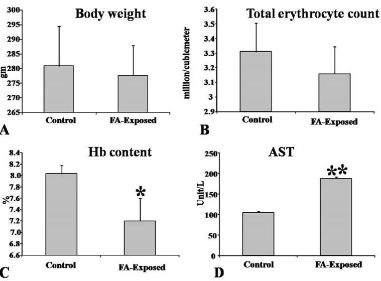
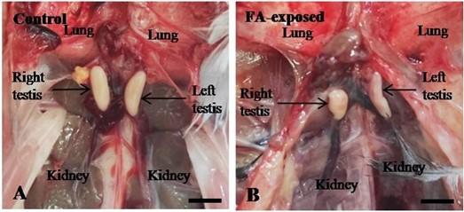
The mean body weights of adult pigeons were not changed significantly in FA-exposed pigeons in comparison with control pigeons (Figure 1A). The color, size and shape of both testes (right and left) were normal in control pigeons (Figure 2A). However, in FA-exposed pigeon, the testes showed uneven shape with pin point hemorrhage on their surface (Figure 2B). The weight of the testes was not change significantly in FA-exposed and control pigeons (data not shown).
Effects of FA on hematological parameters of pigeons
Total erythrocyte count showed no significant change in FA-exposed pigeons in comparison with control pigeons (Figure 1B). The hemoglobin concentration was significantly decreased in FA-exposed pigeons in comparison with control pigeons (Figure 1C). The values of AST were significantly increased in FA-exposed pigeons in comparison with control pigeons (Figure 1D), an indicative of liver injury and disturbance of body homoeostasis.
Effects of FA on histoarchitecture of testes of pigeons
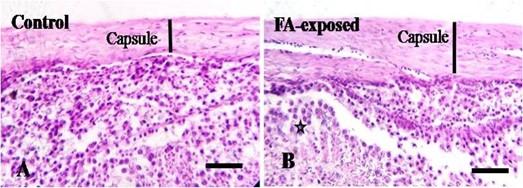
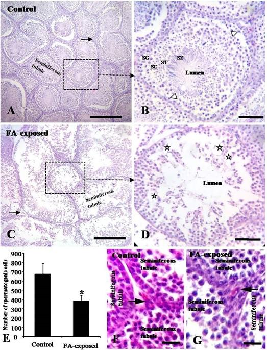
In HE-stained sections, regular histological structure without abnormalities was seen in testes of control pigeons (Figures 3A, 4A-B). The testis was surrounded by a capsule, which was composed mainly of dense collagenous fibrous connective tissue (Figure 3A). The structural components of the testis were the seminiferous tubules and interstitial tissues (Figure 4A-B, F). Seminiferous tubules are the structural and functional unit of testes. The sertoli cells and the spermatogenic cells lined the seminiferous tubules. The different stages of spermatogenic cells were found in several layers, namely, the spermatogonia, spermatocytes, spermatids and finally mature spermatozoa (Figure 4A-B). The spermatogonia were rounded cells with rounded nuclei; and sertoli cells were tall cells with oval nuclei (Figure 4B). The interstitial tissues were narrow and showed clusters of Leydig cells (Figure 4A-B, F).
In FA-exposed pigeons, the FA-induced testicular changes were characterized by thickened capsule and degeneration of spermatogenic cells in the seminiferous tubules (Figure 3B). Irregular arrangement of spermatogenic cells in the seminiferous tubules of FA-exposed pigeons were seen (Figure 4D). Lumen of the seminiferous tubules became large and number of spermatogenic cells particularly; secondary spermatocytes and spermatid were reduced (Figure 4C-D). Primary spermatocytes were found separated from spermatogonia. The number of spermatogenic cells was significantly decreased in seminiferous tubules of FA-exposed pigeons in comparison with control pigeons (Figure 4E). The interstitial cells or Leydig cells were localized as cord or cluster between or among the seminiferous tubules (Figure 4 A-D, F-G). No changes were seen in the Leydig cells of both control and FA-exposed pigeons (Figure 4 F-G).
DISCUSSION
In the present study, the value of serum AST level was significantly increased and the hemoglobin concentration was significantly decreased in FA-exposed pigeons in comparison with control pigeons. The total erythrocyte count showed decreasing tendency in FA-exposed pigeons. These results suggested liver injury and imbalance of body homoeostasis caused by FA exposure. No reports are available in birds, however serum enzyme AST, ALT was significantly increased in FA-exposed mice (oral, 5mg/kg body weight) [22]. FA treatment (oral, 10mg/kg body weight) resulted in significant decreases in RBCs, hemoglobin, total WBC and lymphocytes in mice [23].
FA exposure through feed induced a significant reduction of spermatogenic cells and widening of the lumen of the seminiferous tubules in the testes of pigeons. There was no available literature on FA effect on testicular tissues of avian species. However, the present results were consistent with that of other formaldehyde exposure research on male rats and mice although the dosage, duration and route of administrations of FA were different [13, 21, 24-27]. Low levels of formalin (10 ml/kg feed), when fed to Japanese quails (Coturnix coturnix japonica), decreased testicular weight and diameter of seminiferous tubules [21]. Intraperitoneal administration of FA with dosages of 0.2, 2 and 20 mg/kg can cause degeneration and necrosis of the secondary spermatocytes, spermatogenic cells and spermatozoids [15]. FA vapor (10 mg/m3) for two weeks’ exposure, caused atrophy of the seminiferous tubules, a decrease in the number of spermatogenic cells and disorganization of the seminiferous epithelial cells of male rats [26]. FA-induced change in rat testes are characterized by thickened capsule, spermatogenesis arrest, a decrease in the number of spermatogenic cells and atrophy of seminiferous tubules in rats [28,29], which were reproduced in the present study following FA contaminated feed exposure in testes of adult pigeons.
CONCLUSIONS
The present findings revealed that the toxic feed contaminant, FA, has detrimental effects on the testes of adult pigeons. FA was found to cause degeneration of spermatogenic cells, separation of primary spermatocyte from spermatogonia and irregular arrangement of spermatogenic cells in the seminiferous tubules, these histological changes indicated that it might affect the reproduction of birds. Therefore, it is very alarming if the human being exposed to this toxic food contaminant, it may cause reproductive failure.
ACKNOWLEDGEMENT
The authors thank Mr. Md. Ahasan Habib for chemical preparation and their exposure in birds through feed. This work was supported by the Ministry of Science and Technology (MoST), Government of the Peoples Republic of Bangladesh (No: 39.00.0000.009.14.004.19/ BS-74/85, 2018-19 to MRK), by the Bangladesh Agricultural University and University Grant Commission of Bangladesh (No. 2018/556/AU-GC to MRK) and by the Ministry of Education, Grant for Advance Research in Education (GARE), BANBEIS, Grant Number LS2018773 to MRK.
AUTHOR CONTRIBUTIONS
The experiment was designed by MRK and MP. MRK, AK and IH undertook the experiment; MRK and IH interpreted the results putting efforts on statistical analysis with AK; MRK and IH wrote up the draft. MRK, MP and AIA checked the manuscript critically. All authors read and agreed on the final version of the manuscript.
CONFLICTS OF INTEREST
Authors declared that they have no conflict of interest.Authors declared that they have no conflict of interest.
References
- [1]Rahman M. Sultan Z, Rahman M, Rashid M. Food Adulteration: a serious public health concern in Bangladesh. Bangladesh Pharmaceutical Journal. 2015;18:1
- [2]Nunes MCN. Impact of environmental conditions on fruit and vegetable quality. Stewart Postharvest Review 2008; 4:4
- [3]Karimov K, Dadazhanov SH, Gil’dieva MS. Rat reproductive cells as biological indicators of the effect of environmental factors. Morfologiia (Saint Petersburg, Russia). 2003; 123(1):69-71.
- [4]Handagama C and Ariyaratne S. Differentiation of the adult Leydig cell population in the postnatal testis. Biology of Reproduction. 2001; 65: 660-671.
- [5]Formaldehyde, 2-butoxyethanol and 1-tert-butoxypropan-2-ol. Monographs on the evaluation of carcinogenic risks to humans. 88, 1- 478 (2006).
- [6]Tang X, Bai Y, Duong A, Smith MT, Li L, Zhang L. Formaldehyde in China: production, consumption, exposure levels, and health effects. Environment International. 2009; 35: 1210–1224.
- [7]Brown HR. FDA approved use of formaldehyde in poultry feed. Food Stuffs. 1996; 68. 15
- [8]Duncan MS and Adams AW. Effects of a chemical additive and of formaldehyde-gas fumigation on Salmonella in poultry feeds. Poultry Science. 1972; 51: 797–802.
- [9]Bugarski D, Handzic R, Sivcevic A, Stanic I. Effect of feeding regime and formaldehyde-treated soyabean on the productivity of dairy cows. Veterinaria Sarajevo. 1990; 39: 21–30.
- [10]McAllister T, Beauchemin K, McClelland L, Cheng, K. Effect of formaldehyde-treated barley or escape protein on nutrient digestibility, growth and carcass traits of feedlot lambs. Canadian Journal of Animal Science. 1992; 72: 309–316.
- [11]Ricke SC, Richardson K, Dittoe DK. Formaldehydes in feed and their potential interaction with the poultry gastrointestinal tract microbial community–A review. Fronters in Veterinary Science. 2019; 6: 188. doi: 3389/fvets.2019.00188
- [12]Duong A, Steinmaus C, McHale CM, Vaughan CP, Zhan L. Reproductive and developmental toxicity of formaldehyde: A systematic review. Mutation Research. 2011; 118-138.
- [13]Zhou D, Zhang J, Wang H. Assessment of the potential reproductive toxicity of long-term exposure of adult male rats to low-dose formaldehyde. Toxicology and Industrial Health. 2011; 27(7):591-5988.
- [14]Zahra T, Parviz T, Simin F, Mehdi T. Effect of formaldehyde injection in mice on testis function. International Journal of Pharmacology. 2007; 3(5):421-424.
- [15]Tang M, Xie Y, Yi Y, Wang W. Effects of formaldehyde on germ cells of male mice. Wei sheng yanjiu= Journal of hygiene research. 2003; 32(6):544-548.
- [16]Chung WG, Yu IJ, Park CS, Lee KH, Roh HK, Cha YN. Decreased formation of ethoxyacetic acid from ethylene glycol monoethyl ether and reduced atrophy of testes in male rats upon combined administration with toluene and xylene. Toxicology Letters. 1999; 104(1-2):143-150.
- [17]Lemasters GK, Olsen DM, Yiin JH, Lockey JE, Shukla R, Selevan SG, Schrader SM, Toth GP, Evenson DP, Huszar GB. Male reproductive effects of solvent and fuel exposure during aircraft maintenance. Reproductive Toxicology. 1999; 13(3):155-166.
- [18]Özen OA, Akpolat N, Songur A, Kuş İ, Zararsiz İ, Özaçmak VH, Sarsilmaz M. Effect of formaldehyde inhalation on Hsp70 in seminiferous tubules of rat testes: an immunohistochemical study. Toxicology and Industrial Health. 2005; 21(9):249-254.
- [19]Golalipour MJ, Azarhoush R, Ghafari S, Gharravi AM, Fazeli SA, Davarian A. Formaldehyde exposure induces histopathological and morphometric changes in the rat testis. Folia Morphologica. 2007; 66(3):167-171.
- [20]Babar AM, Khan MZ, Ahmed S, Khan A, Bachaya, Anwar MI. Toxico-pathological effects of formalin (37% formaldehyde) feeding in broiler chicks. Pakistan Veterinary Journal. 2001; 21:13-16.
- [21]Anwar MI, Khan MZ, Muhammad G, Bachaya A and Babar MA. Effects of dietary formalin on the health and testicular pathology of male Japanese quails (Coturnix coturnix japonica). Veterinary and Human Toxicology.2001; 43: 330–333.
- [22]Afrin M, Amin T, Karim, MR, Islam MR. Effects of formaldehyde intoxication on liver of Swiss albino mice. IOSR Journal of Agriculture and Veterinary Science. 2016; 9: 76-81.
- [23]Abd-Elhakim YM, Amany M, & Wafaa M. Hemato-immunologic impact of sub chronic exposure to melamine and/or formaldehyde in mice, Journal of Immunotoxicology. 2016; 13(5):713-722.
- [24]Majumder PK, Kumar VL. Inhibitory effects of formaldehyde on the reproductive system of male rats. Indian Journal of Physiology and Pharmacology. 1995; 39(1):80-82.
- [25]Shah BM, Vachharajani KD, Chinoy NJ and Roy Chowdhary A. Formaldehyde-induced changes in testicular tissues of rats. Journal of Reproductive Biology and Comparative Endocrinology.1992; 7: 42–52.
- [26]Zhou DX, Qiu SD, Wang ZY and Zhang J. Effect of tail-suspension on the reproduction of adult male rats. Zhonghua Nan KeXueZaZhi. 2006a; 12: 326–329.
- [27]Zhou DX, Qiu SD, Zhang J, Tian H and Wang HX. The protective effect of vitamin E against oxidative damage caused by formaldehyde in the testes of adult rats. Asian Journal of Androoglogy.2006b; 8: 584–588.
- [28]Hegazy AA, Elsayed NE, Ahmad MM, Omar NM. Effect of formaldehyde on rat testis structure. Academia Anatmica International. 2017; 3(2):15-23.
- [29]Chowdhury AR, Gautam AK, Patel KG, Trivedi HS. Steroidogenic inhibition in testicular tissue of formaldehyde exposed rats. Indian Journal of Physiology and Pharmacology. 1992; 36: 162.