Antagonistic activity of Exiguobacterium indicum LIS01 isolated from sediment
Abstract
The bacterial resistance over antibiotics has gained significant concern worldwide for the last few decades. This study aimed to isolate and characterize antibiotic producing bacteria from different water sediment sources in Bangladesh. A total of 9 samples were collected from three different sediment sources from Gopalganj, Bangladesh. Bacterial isolates grown in nutrient agar with cycloheximide were tested for antagonistic activity following disc-diffusion assay. Based on preliminary careening, three isolates namely LIS01, LIS02 and LIS03 showed antibacterial activity against six clinical isolates: E. coli, Salmonella sp., Shigella sp., Klebsiella pneumoniae, Streptococcus sp., and Staphylococcus sp. The isolate LIS01 with the highest (P<0.05) zone of inhibition was characterized using 16S rRNA gene sequencing. According to phylogenetic analysis, the isolate was identified as Exiguobacterium indicum with more than 99.7% sequence similarity to previously characterized strains. The results of our study are very promising considering the antibiotic resistant bacteria and could be helpful in treating diseases associated with drug failure caused.
INTRODUCTION
The emergence of drug resistance in clinical isolates causes severe health alarm globally. In developing countries with poor health and hygiene, the problem becomes worse day by day [1,2]. In addition, environmental pollution, mostly by industrial and medical waste generates super bags through horizontal transfer of antibiotic resistant genes from pathogenic microbes into non-pathogenic isolates [3,4]. This cycle continues and gives rise to numerous superbugs, making the treatment process difficult and complicated. In Bangladesh, most recent studies revealed alarming drug resistant patterns in clinical isolates where bacteria gained complete resistance against third generation antibiotics (ceftriaxone, cephalosporin and ciprofloxacin) [4–8]. Considering the side effects, environmental impacts, and sustainability, finding an alternative source of antibiotics has still remained the utmost priority in biological science and natural product research.
Environment is considered as the natural reservoir of a vast range of antagonistic bacteria with therapeutic applications [9–11]. Among the environmental bacteria, Streptomyces, Bacillus and Pseudomonas species are the most important bacteria reported to produce broad spectrum antibiotics having agricultural and medical applications [4,12–14]. Although previous studies demonstrated the antagonistic activity of these isolates against drug resistant clinical isolates [15–20], however re-emergence of virulent strains and ever changing counter mechanisms of bacteria against drugs compelling researchers to find new antagonistic bacteria with potent anti-microbial activity. Bangladesh has a diverse land area, ocean, rivers, hills and mangrove forest that are reported as the most potent sources of antagonistic bacteria [21–23]. Especially the soil and sediment bacteria due to their diversity and distribution produce a wide range of bioactive metabolites and antimicrobial proteins [24–26]. In this backdrop, the present research aimed to characterize new, safe, eco-friendly, and potent antagonistic bacteria from the river sediment of Bangladesh and evaluate the effects against a broad range of clinical pathogens.
MATERIALS AND METHODS
Collection of bacterial isolates
Bacterial samples were collected from three different places of Gopalganj (23.0130° N, 89.8224° E), Bangladesh. The sampling points are Madhumoti river (latitude: 22° 52′ 47″ N, longitude: 89° 54′ 45″ E), Chandar Beel (23.2632° N, 89.9066° E) and Borni baor (22.9602° N, 89.8523° E). A total of 9 sediment samples (n = 3) were collected from at least 15 cm of depth with a sterile inoculating spoon during March and April 2019, placed in sterile plastic zip-lock bag and transported immediately (< 1 h) to the Biotechnology and Genetic Engineering-laboratory of the Bangabandhu Sheikh Mujibur Rahman Science and Technology University, Bangladesh, by maintaining cold chain with gel ice. Then, the samples were air dried according to methods described by Hossain and Rahman [23].
Culture conditions and biochemical characterization
For the isolation of bacteria, 10- fold serial dilution method was performed for 9 samples with phosphate buffered saline (PBS, pH 7.2). Sample from each dilution (104-107) was then streaked on a nutrient agar (NA) plate amended with cycloheximide (100 mg/ml) to prevent fungal growth and incubated at 37°C for 24 h [23]. Isolated colonies were then sub-cultured into NA plate and incubated overnight at 37°C [27]. Primary biochemical characterization was performed according to Bergey’s manual for the identification of bacteria [28]. Further biochemical characterization was performed following methods for identification of Bacillus and Exiguobacterium species [29,30].
Evaluation of antagonistic activity
The antagonistic activity of bacterial isolates was measured by cross streak method described by Ran et al., 2012 [31]. A pure culture of each isolate was first grown on the nutrient broth (NB) and incubated for 24 h at 37°C. From the NB broth, 50 ml of culture was poured in 6 mm circular Whatman’s filter paper, dried at room temperature, followed by disc-preparation using a punching machine [26]. Six clinical isolates namely E. coli, Salmonella sp., Shigella sp., Klebsiella pneumoniae, Streptococcus sp., and Staphylococcus sp. collected from Gopalganj medical college and hospital were used to evaluate the antagonistic activity of cultured isolates. The disc-diffusion assay was performed following methods described by Ran et al., 2020. These clinical isolates were grown on nutrient broth (NA) culture and incubated overnight at 37 °C. Then, 50 ml of bacterial culture was grown in the nutrient agar (NA) by spread plate method [26]. Then previously prepared filter paper discs of bacterial isolates were carefully placed onto the culture plate skeptically under the biological safety cabinet. In the present study, we analyzed 12 h, 18 h, and 24 h of bacterial culture for the inhibition assay. After 24 h of incubation, the zone of inhibition was measured according to the method described earlier [26]. Each experiment was performed as triplicate.
Characterization of potential antagonistic bacteria
The isolate with highest antagonistic activity was selected for further study. It was sub-cultured again on nutrient broth for molecular characterization using 16S rRNA sequencing. The overnight broth culture was used for bacterial genomic DNA extraction using Genomic DNA Mini Prep Kit (Bio Basic Inc., Ontario, Canada) according to manufacturer’s instructions. The extracted DNA was checked for quality and quantity in 1% agarose gel using lambda (k) DNA marker and Nanodrop spectrophotometer (Thermo Fisher Scientific, Waltham, USA) as a ratio of DNA-protein absorbance. The DNA was then stored at -20°C until further use.
PCR amplification of bacteria
The PCR was performed in 25 µl master reaction mixtures containing template 1.5 µl DNA (genomic DNA of bacteria), 2.5 µl of 25 mM MgCl2, 2.5µl of 10x colorless reaction buffer, 1.0 µl concentration of deoxynucleotide triphosphate (dNTP), 1 µl of each of universal forward (27 F, 5′- AGA GTT TGA TCM TGG CTC AG -3′) and reverse primer (1391 R, 5′- GGT TAC CTT GTT ACG ACT T -3′) for bacteria, 0.5µl Taq DNA polymerase and 15 µL of nuclease-free water. A total of 35 cycles of amplification reactions were carried out in a MultiGene gradient thermal cycler (Labnet International Inc., USA) under following conditions; an initial denaturation step at 95°C for 5 min followed by a second denaturation step at 95°C for 40 sec, annealing for 1.0 min at 55°C, an elongation at 72°C for 2.0 min, and a final extension step of 72°C for 10 min [32].
Sequencing and analysis of 16s rDNA
The PCR product was purified using the PureLink PCR purification kit (ThermoFisher Scientific, USA) according to the manufacturer’s guidelines. The purified PCR product was then sent to Invent Technologies Limited, Dhaka, for 16s rDNA sequencing. The sequences were then edited and assembled in Geneious software (vR11.1) [33] and before further phylogenetic analysis using MEGA 7.0 [34]. After primary nucleotide BLAST (BLASTn) in NCBI, multiple sequence alignment was performed as ClustalW in MEGA 7.0. The phylogenetic tree was constructed using neighbour-joining method with 1000 bootstrap replicates, taking Streptomyces sp. as the out-group strain. The nucleotide sequence of the bacteria is currently available at NCBI GenBank database under the accession number of MT742616.1.
Statistical analysis
One-way ANOVA with Tukey’s post hoc test was applied to compare the mean values of antagonistic activity for three isolates (LIS01, LIS02 and LIS03). Alpha level of 0.05 was considered as statistically significant.
RESULTS
Isolation of bacteria
Based on morphological and biochemical tests (Table 1), three isolates primarily identified as Bacilli. However, isolate LIS01 showed differences in biochemical characteristics compared to LIS02 and LIS03, indicating a different bacteria strain.
Table 1. Biochemical characterization of isolates.
Antagonistic activity of E. indicum LSA01
In primary screening, the isolate E. indicum LSA01 showed significantly higher (P<0.05) antagonistic activity compared to other two isolates (LIS02 and LIS03) (Figure 1). The E. indicum LSA01 showed potential inhibitory effects against all the six tested clinical isolates in the in vitro plate assay. Study results revealed that the overnight culture (24 h) was most effective (P<0.05) against tested isolates, followed by 18 h and 12 h, respectively. The highest zone of inhibition with E. indicum LAS01 was 16.3 mm for E. coli, followed by 15.3 mm for Shigella sp., 12.3 mm for Salmonella sp., and Streptococcus sp., and 11.7 mm for Staphylococcus sp., and 10.7 mm for K. pneumoniae, respectively (Table 2).
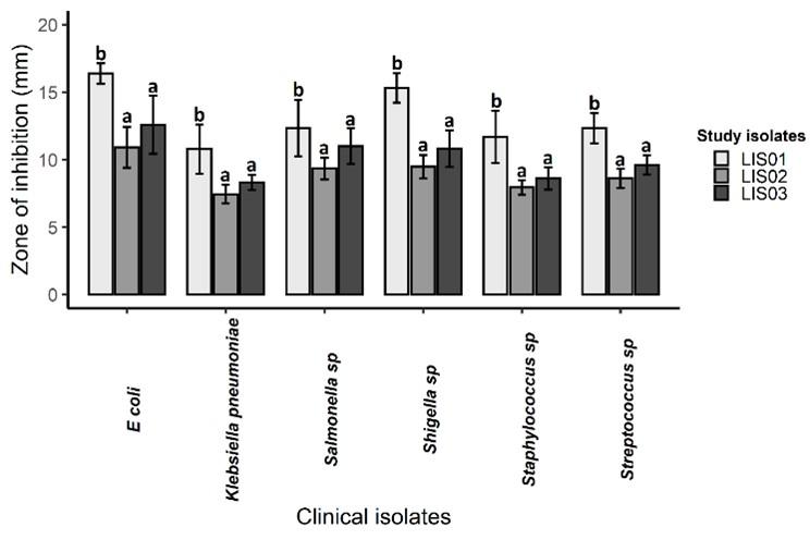
Table 2. Antagonistic activity (mean ± SE) of E. indicum LSA01 against six clinical isolates.
Identification of bacteria and phylogenetic analysis
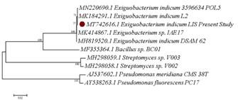
The modified tests for the classification of Exiguobacterium genus [29] confirmed the highest antibiotic producing isolate LIS01 as E. indicum. The NCBI nucleotide BLAST (BLASTn) and phylogenetic tree analysis using 16S rRNA data confirmed the species and relationship of LIS01 to previously identified E. indicum (Figure 2). Neighbour-joining tree revealed close clustering of E. indicum LSA01 with other previously identified E. indicum including strain isolated from lake, river, and marine water in India. The study isolates also upheld close relationship to Bacillus species, however distant to Streptomyces species, as shown in phylogenetic tree (Figure 2).
DISCUSSION
Emergence of antibiotic resistant bacteria compelled scientists to find new and effective drugs to prevent and treat many diseases of human. Compared to other sources, biocontrol agents are effective, safe and eco-friendly, and therefore used in novel drug developments since the discovery of penicillin [35]. Nature represents the most significant resource for the biologically active compounds that can be used as antibiotics to fight against bacterial diseases [36]. In this study, we have identified an Exiguobacterium species that found to have potent inhibitory activity against six MDR clinical isolates. The isolate was confirmed as E. indicum by biochemical and 16S rRNA based sequencing. The 16S rRNA method has been considered as gold standard for the identification of bacterial species [37,38]. Considering the drug resistant patterns in clinical isolates, the E. indicum species could be used as potential antibiotic candidate for treating diseases associated with these six bacteria.
The isolates were screened for antibiotics production through disc diffusion in the in vitro plate assay. This is an agreement with some previous literatures that used the same methods to screen prospective antibiotic producing isolates [26,39,40]. In present study, E. indicum strongly inhibit E. coli and Shigella spp., indicating its higher efficacies against food-borne pathogens. However, the activity was significantly lower for Staphylococcus and Klebsiella, two most drug resistant clinical bacteria characterized from urinary tract infected (UTI) patients in Bangladesh [41–43]. These results are inconsistent with some previous studies where the higher inhibitory activity of Streptomyces and Bacillus species was recorded against E. coli compared to Streptococcus, Staphylococcus and Klebsiella species [17,44,45]. In recent studies, complete drug resistant Staphylococcus and Klebsiella isolates have been identified from patients with UTI revealing the severity of drug incompetence in Bangladesh [41].
The phylogenetic data of E. indicum revealed a close relationship of present study isolate with other antagonistic Exiguobacterium species isolated from soil, water and sediments [41,46-47]. Out of these close neighbours, E. indicum POL5 had highest similarity score to present study isolate that is reported as potential inhibitor of pathogenic bacteria. To compare with other potential antagonistic strains, we found that present study isolate was distinctly different from Bacillus sp. BC01 and Streptomyces species. Therefore, it is quite clear that, present study bacteria are a new strain of E. indicum with promising antagonistic activity against clinical isolates.
After investigating the results, the present study strain could be used as potential antibiotic candidate after purification and necessary strain development processes. However, considering the limited resources and financial constraints, those steps were beyond our study objectives. In addition, the number of clinical isolates (six species) tested is not big enough to make a conclusion about the broad-spectrum antagonistic activity of present study bacteria. Therefore, inhibitory activity should be measured with big numbers and greater volume of clinical samples. Further genome sequencing and functional annotations are required to identify the candidate genes associated with antagonistic activity.
ACKNOWLEDGEMENT
None.
AUTHOR CONTRIBUTIONS
AKL designed the study, performed laboratory assays, analyzed the data, and prepared the draft manuscript. MSA and FHM collected samples and performed inhibitory assays. AAJ analyzed the data and reviewed the manuscript. All authors approved the final manuscript.
CONFLICTS OF INTEREST
There is no conflict of interest among the authors.
References
- [1]World Health Organization. WHO global strategy for containment of antimicrobial resistance, World Health Organisatin. WHO Glob Strateg Contain Antimicrob Resist. 2001;1–105.
- [2]Ayukekbong JA, Ntemgwa M, Atabe AN. The threat of antimicrobial resistance in developing countries: Causes and control strategies. Antimicrob Resist Infect Control. 2017;6(1):1–8.
- [3]Chowdhury FFK, Acharjee M, Noor R. Maintenance of Environmental Sustainability Through Microbiological Study of Pharmaceutical Solid Wastes. Clean – Soil, Air, Water. 2016;44(3):309–316.
- [4]Moges F, Endris M, Belyhun Y, Worku W. Isolation and characterization of multiple drug resistance bacterial pathogens from waste water in hospital and non-hospital environments, Northwest Ethiopia. BMC Res Notes. 2014;7(1):1–6.
- [5]Shamsuzzaman A, Paul SK, Mahmud MC, Musa A, Hossain MA. Emerging antimicrobial resistance amongst common bacterial pathogens in Mymensingh Medical College Hospital. Bangladesh J Med Microbiol. 2016;1(1):4–9.
- [6]Rashid A, Chowdhury A, Rahman SH, Begum SA, Muazzam N. Infections by Pseudomonas aeruginosa and antibiotic resistance pattern of the isolates from Dhaka Medical College Hospital. Bangladesh J Med Microbiol. 2016;1(2):48–51.
- [7]Siddiqua M, Alam AN, Akter S, Ferdousi RS. Antibiotic resistance pattern of bacteria causing urinary tract infection in a private medical college hospital, Dhaka. Bangladesh J of Med Sci. 2015;16(01):42–47.
- [8]Ahmed I, Rabbi MB, Sultana S. Antibiotic resistance in Bangladesh: A systematic review. Int J Infect Dis. 2019;80(January):54–61.
- [9]Cycoń M, Mrozik A, Piotrowska-Seget Z. Antibiotics in the soil environment—degradation and their impact on microbial activity and diversity. Front Microbiol. 2019;10:Article 338.
- [10]Feichtmayer J, Deng L, Griebler C. Antagonistic microbial interactions: Contributions and potential applications for controlling pathogens in the aquatic systems. Front Microbiol. 2017;8(11):1–14.
- [11]Hatosy SM, Martiny AC. The ocean as a global reservoir of antibiotic resistance genes. Appl Environ Microbiol. 2015;81(21):7593–7599.
- [12]Naturally Occurring Antimicrobial Drugs : Antibiotics. 2020;13.3B: 1–2.
- [13]Rafiq A, Khan SA, Akbar A, Shafi M, Ali I, Ur Rehman F, et al. Isolation And identification of antibiotic producing microorganisms from soil. Int J Pharm Sci Res. 2015;9(3):1002–1011.
- [14]Cesa-Luna C, Baez A, Quintero-Hernández V, De La Cruz-Enríquez J, Castañeda-Antonio MD, Muñoz-Rojas J. The importance of antimicrobial compounds produced by beneficial bacteria on the biocontrol of phytopathogens. Acta Biol Colomb. 2020;25(1):140–154.
- [15]Hasan FB, Reza M, Masud HA Al, Uddin MK, Uddin MS. Preliminary characterization and inhibitory activity of bacteriocin like substances from Lactobacillus casei against multi-drug resistant bacteria. Bangladesh J Microbiol. 2019;36(1):1–6.
- [16]Lagadinou M, Onisor MO, Rigas A, Musetescu DV, Gkentzi D, Assimakopoulos SF, et al. Antimicrobial properties on non-antibiotic drugs in the era of increased bacterial resistance. Antibiotics. 2020;9(3):1–12.
- [17]Monte J, Abreu AC, Borges A, Simões LC, Simões M. Antimicrobial activity of selected phytochemicals against Escherichia coli and Staphylococcus aureus and their biofilms. Pathogens. 2014;3(2):473–498.
- [18]Bantawa K, Sah SN, Subba Limbu D, Subba P, Ghimire A. Antibiotic resistance patterns of Staphylococcus aureus, Escherichia coli, Salmonella, Shigella and Vibrio isolated from chicken, pork, buffalo and goat meat in eastern Nepal. BMC Res Notes. 2019;12(1):1–6.
- [19]Chattopadhyay MK, Chakraborty R, Grossart HP, Reddy GS, Jagannadham M V. Antibiotic resistance of bacteria. Biomed Res Int. 2015;2015.
- [20]Hemashenpagam N, Saranya S. Antagonistic activity and antibiotic sensitivity of Lactic acid bacteria from fermented dairy products. Adv Appl Sci Res. 2011;2(4):528–534.
- [21]Rahman MM, Ali ME, Khan AA, Akanda AM, Uddin MK, Hashim U, et al. Isolation, characterization, and identification of biological control agent for potato soft rot in Bangladesh. Sci World J. 2012;2012.
- [22]Rahman MM, Khan AA, Ali ME. Screening of antagonistic bacteria against bacterial wilt of tomato, eggplant and potato in Bangladesh. Int J Agric Biol. 2013;15(5):973–977.
- [23]Hossain MN, Rahman MM. Antagonistic activity of antibiotic producing Streptomyces sp. against fish and human pathogenic bacteria. Brazilian Arch Biol Technol. 2014;57(2):233–237.
- [24]Mandal C, Tabassum T, Shuvo M, Habib A. Biochemical and molecular identification of antibiotic-producing bacteria from waste dumpsite soil. J Adv Biotechnol Exp Ther. 2019;2(3):120.
- [25]Gebreyohannes G, Moges F, Sahile S, Raja N. Isolation and characterization of potential antibiotic producing actinomycetes from water and sediments of Lake Tana, Ethiopia. Asian Pac J Trop Biomed. 2013;3(6):426–435.
- [26]Foysal MJ, Lisa AK. Isolation and characterization of Bacillus sp. strain BC01 from soil displaying potent antagonistic activity against plant and fish pathogenic fungi and bacteria. J Genet Eng Biotechnol. 2018;16(2):387–392.
- [27]Cazorla FM, Romero D, Pérez-García A, Lugtenberg BJJ, Vicente A De, Bloemberg G. Isolation and characterization of antagonistic Bacillus subtilis strains from the avocado rhizoplane displaying biocontrol activity. J Appl Microbiol. 2007;103(5):1950–1959.
- [28]Krieg NR. Identification of Procaryotes. Bergey’s Manual® Syst Bacteriol. 2001;33–38.
- [29]Chaturvedi P, Shivaji S. Exiguobacterium indicum sp. nov., a psychrophilic bacterium from the Hamta glacier of the Himalayan mountain ranges of India. Int J Syst Evol Microbiol. 2006;56(12):2765–2770.
- [30]Wulff EG, Mguni CM, Fels J, Hockenhull J. Biochemical and molecular characterization of Bacillus amyloliquefaciens, B . subtilis and B . pumilus isolates with distinct antagonistic potential against Xanthomonas campestris pv . campestris. 2002;574–584.
- [31]Ran C, Carrias A, Williams MA, Capps N, Dan BCT, Newton JC, et al. Identification of Bacillus strains for biological control of catfish pathogens. PLoS One. 2012;7(9).
- [32]Frank JA, Reich CI, Sharma S, Weisbaum JS, Wilson BA, Olsen GJ. Critical evaluation of two primers commonly used for amplification of bacterial 16S rRNA genes. Appl Environ Microbiol. 2008;74(8):2461–2470.
- [33]Kearse M, Moir R, Wilson A, Stones-Havas S, Cheung M, Sturrock S, et al. Geneious Basic: An integrated and extendable desktop software platform for the organization and analysis of sequence data. Bioinformatics. 2012;28(12):1647–1649.
- [34]Kumar S, Stecher G, Tamura K. MEGA7: Molecular Evolutionary Genetics Analysis version 0 for bigger datasets. Mol Biol Evol. 2016;33(7):1870–1874.
- [35]Barratt BIP, Moran VC, Bigler F, van Lenteren JC. The status of biological control and recommendations for improving uptake for the future. BioControl. 2018;63(1):155–167.
- [36]Bérdy J. Thoughts and facts about antibiotics: Where we are now and where we are heading. J Antibiot (Tokyo). 2012;65(8):385–395.
- [37]Janda JM, Abbott SL. 16S rRNA gene sequencing for bacterial identification in the diagnostic laboratory: Pluses, perils, and pitfalls. J Clin Microbiol. 2007;45(9):2761–2764.
- [38]Clarridge Jill E. Impact of 16S rRNA gene sequence analysis for identification of bacteria on clinical microbiology and infectious diseases. Clin Microbiol Rev. 2004;17(4):840–862.
- [39]Kaur S, Kaur J, Pankaj PP. Isolation and characterization of antibiotic producing microorganisms from soil samples of certain area of Punjab region of India. Int J Pharm Clin Res. 2014;6(4):312–315.
- [40]Moore T. Antagonistic activity of Bacillus bacteria against food-borne pathogens. J Probiotics Heal. 2013;01(03):1–6.
- [41]Mahmudunnabi G, Majlish ANK, Momtaz F, Foysal MJ, Rahman MM, Islam K. Molecular detection and PCR-RFLP analysis using Pst1 and Alu1 of multidrug resistant Klebsiella pneumoniae causing urinary tract infection in women in the eastern part of Bangladesh. J Genet Eng Biotechnol. 2018;16(1):77–82.
- [42]Majlish ANK, Momtaz F, Foysal MJ, Islam K, Alam MJ, Prodhan MSH. Characterization of multi-drug resistant Klebsiella pneumoniae isolates from urinary tract infected-women in Sylhet city, Bangladesh. Malays J Microbiol. 2019;15(6):455–462.
- [43]Sharmin L, Akter S. Bacterial aetiology and antibiotic resistance pattern of community-acquired urinary tract infections in children in a tertiary care hospital in Bangladesh. J Enam Med Coll. 2017;7(3):134–139.
- [44]Kadhum HA, Hasan TH. The study of Bacillus subtils antimicrobial activity on some of the pathological isolates. Int J Drug Deliv Technol. 2019;9(2):193–196.
- [45]Hasan N, Yang H. Factors affecting the composition of the gut microbiota, and its modulation. PeerJ. 2019;2019(8):1–31.
- [46]Kasana RC, Pandey CB. Exiguobacterium: an overview of a versatile genus with potential in industry and agriculture. Crit Rev Biotechnol. 2018;38(1):141–156.
- [47]Selvakumar G, Joshi P, Nazim S, Mishra PK, Kundu S, Gupta HS. Exiguobacterium acetylicum strain 1P (MTCC 8707) a novel bacterial antagonist from the North Western Indian Himalayas. World J Microbiol Biotechnol. 2009;25(1):131–137.