Antibacterial and phytochemical effects of ethanol extracts of Syzygium guineense (Willd.) DC barks and Mangifera indica L seeds
Abstract
Bacterial infectious diseases account for thousands of deaths worldwide. Despite their side effects, synthetic antibiotics are currently utilized to treat bacterial infections. There has been an effort to identify alternative medicines of plant origin. Thus, the current study determined in vitro antibacterial activities of Syzygium guineense barks and seeds of Mangifera indica ethanol extracts, as well as their phytochemical profile. The tested bacteria included Salmonella typhi, Staphylococcus aureus, Bacillus subtilis, and Escherichia coli. Plant samples were collected from Morogoro region, Tanzania and transferred to Kenyatta University for preparation and extraction. In vitro antibacterial activities were determined by disk diffusion, MIC, and MBC methods. Selected phytochemicals of ethanol extracts were determined qualitatively. The highest antibacterial effects were observed in M. indica extract against B. subtilis, S. aureus, E. coli and S. typhi with zones of inhibition of 20.00 mm, 18.00 mm, 17.67 mm, and 15.67 mm, respectively. Antibacterial effects observed in S. guineense extract against B. subtilis, S. aureus, S. typhi and E. coli produced zones of inhibition of 15.00 mm, 14.33 mm, 10.67 mm, and 9.33 mm, respectively. The extracts showed better antibacterial effects against Gram positive bacteria than Gram negative bacteria. Qualitative phytochemical analysis of the ethanol extracts revealed alkaloids, quinones, flavonoids, phenolics, saponins, steroids, terpenoids, tannins, and glycosides. This study indicates that the ethanol extracts of the plants could be used to develop alternative remedies for treating bacterial infections. The study also suggests that the plant extracts should be subjected to in vivo studies.
INTRODUCTION
Medicinal plants have been used to prevent and manage human diseases since time immemorial and are considered safe, eco-friendly, and locally available [1, 2]. Plants produce phytochemicals that defend them against pathogenic microbes [3]. These phytochemicals have shown benefits in managing diseases like bacterial infections, cancer, malaria, sickle cell anemia, infertility, obesity, and diabetes [4, 5]. Medicinal plants produce different kinds of phytocompounds like phenols, flavonoids, tannins, alkaloids, saponins, and terpenes, which possess antimicrobial activities against many pathogens [3]. Use of medicinal plants provides an alternative remedy to alleviate several ailments brought by bacterial infections in humans.
Bacteria are the most common microorganism that cause majority of infectious diseases responsible for high morbidity and death worldwide [6]. Global trends reported that more than four million deaths occur annually due to bacterial infections, forty-two percent of global deaths occur in Africa [7]. Bacterial infections diseases are conventionally treated with antibiotics, however the undesirable side effect such as causing inflammation, haemolytic anaemia, nausea, rashes, vomiting, and hypersensitivity reactions [8], and increase of multidrug resistant bacteria to antibiotics has made it difficult to manage these infections [6]. The consequences of antibiotic resistance and side effects have accelerated research on using medicinal plants as alternative medicines to fight bacterial infections [9].
M. indica belongs to Anacardiaceae family [10], and used locally to treat diseases like urinary tract infections, gynaecological diseases, diabetes, diarrhea, malaria, Asthma, cough, and toothache [11]. S. guineense is a large tree in the family Myrtaceae widespread in Africa [12]. The roots and stem bark infusions of S. guineense are used by Tanzanian communities to treat typhoid fever, stomachache, diarrhea, diabetes mellitus and as anthelmintic [13].
In Tanzania, medicinal plants are used locally as remedy agents for a number of diseases since they are relatively cheaper, easily available and claimed to be safe and effective [14]. However, there is still a gap confirming the scientific validation of many plant species with medicinal properties from Tanzanian plants. Tanzania has endowed with over 10,000 plant species and a quarter of them are medicinal plants used by people but not all of them have been evaluated scientifically [4]. S. guineense and M. indica are among herbal plants used in folklore medicine by local communities in Morogoro, Tanzania for treating bacterial ailments [9]. However, their antibacterial activities have not been scientifically validated [5]. Therefore, this study intended to determine the phytochemical profile and scientifically validate the antibacterial activities of ethanol extracts of S. guineense and M. indica grown in Tanzania.
MATERIALS AND METHODS
Collection and preparation of medicinal plants
Barks of S. guineense and seeds of M. indica, were collected from Morogoro region in Tanzania with the global positing system (GPS) coordinates of 6° 49' 39.9216'' S and 37° 39' 32.8104'' E. Plant samples were brought to Kenyatta University, for preparation and extraction. A plant taxonomist identified the plants and voucher specimens stored at Kenyatta University herbarium. Plant samples were air dried under shade, grounded into powder using a mechanical mill and stored in air-tight containers until use.
Extraction of plant samples
Two-hundred grams of dry powdered plant samples were separately added to 600 ml of cold ethanol and incubated for forty-eight hours. The resulting mixtures were decanted and filtered using Whatman number 1 filter paper and then concentrated at 40°C using a rotary evaporator. The extracts were placed in airtight containers and stored 4°C until use [15].
Antibacterial assays
Test microbes
Bacteria utilized for this study included Salmonella typhi, Bacillus subtilis, Staphylococcus aureus and Escherichia coli. Cultures of the bacteria were obtained from biochemistry, microbiology and biotechnology laboratories and cultures were maintained on nutrient agar medium.
Preparation of McFarland turbidity standard
The reference stock (McFarland Turbidity Standard) for adjusting bacteria turbidity was made by mixing 9.95ml of 1% sulfuric acid and 0.5ml of 1.2% barium chloride dihydrate in a 10ml sterile test tube which was shaken to maintain the suspension, closed tightly and stored at 25±2°C awaiting use [16].
Innocula suspension preparation
The inoculum suspension was made following a protocol published by Debalke et al. [17]. For each bacteria strain, 4-5 colonies were chosen using a sterile wire-loop and then placed in a sterile test tube with 2ml of normal saline. Then, they were vortexed thoroughly, modified to 0.5 McFarland standards and used within 15 minutes after preparation.
Media preparation
The media used for culture were Muller Hinton Agar (MHA), Muller Hinton Broth (MHB) and nutrient agar media. The media were prepared following the manufacturer’s directions, where 38 grams of MHA and 21 grams of MHB were each added to one liter of distilled water, heated to ensure complete dissolution of the medium followed by autoclaving for 15 minutes at 121°C then cooled to 45℃ and dispensed into sterilized plastic petri dishes to an even depth in a lamina flow chamber. They were then labelled and stored at 2-8℃, awaiting use. The nutrient agar and broth were prepared similarly to MHA and MHB.
Preparation of impregnated paper disc and plant extract dilution
Paper discs with a diameter of 6mm, were punched from Whatman filter paper No 1, after which they were autoclaved for 15 minutes at 121℃. The discs were impregnated with different extract concentrations, positive and negative controls, and left to dry in a lamina airflow for 30 minutes at room temperature before use [15].
Extracts concentration preparation
A 1000 mg/ml stock preparation was made for each plant extract by adding 1 gram of the extract to 1 milliliter of 10% dimethyl sulfoxide (DMSO). Two-fold successive dilutions were made by adding 50µl of the 1000mg/ml starting concentration to 500 microliters of DMSO in the first tube to give 500mg/ml. Further two-fold dilution were done to obtain concentration of 250mg/ml followed by 125, 62.5, 31.25 and 15.625mg/ml.
Antibacterial sensitivity test using disc diffusion
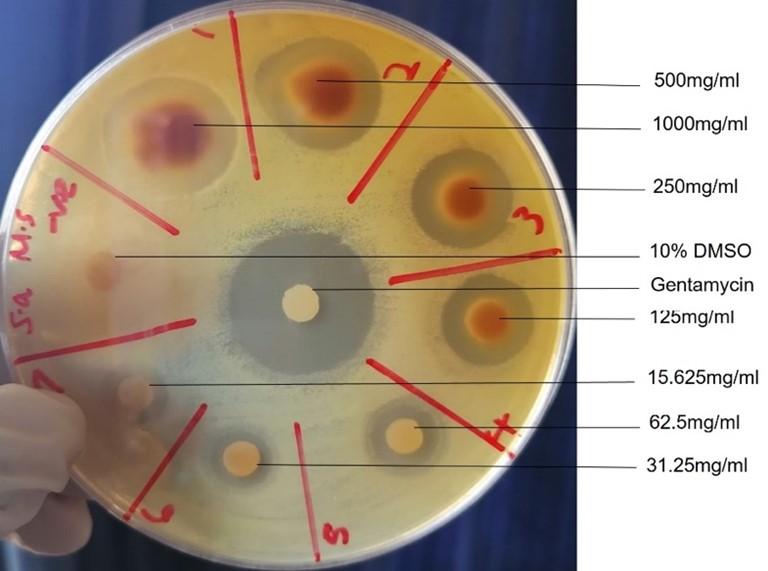
This was done in triplicates using agar diffusion method according to Hudzicki [16]. A sterile cotton swab was dipped into inoculum tube and MHA plate was inoculated by closely streaking the swab to ensure equal distribution of inoculum. Then, the plates were dried in a lamina flow at room temperature for 5 minutes. Paper discs impregnated with the plant’s extracts at various doses were gently placed on the surface of MHA using sterilized forceps. Every plate had 7 discs impregnated with plant extracts, one with 10% DMSO (negative control) and one with 50µg/ml gentamicin (positive control), which were placed equidistant from each other. The plates were maintained at 37 degrees Celsius overnight, after which the diameter of the inhibition zone was determined and presented in millimeters. Clear zones around the discs indicated the ability to inhibit bacterial growth [16].
Determination of minimum inhibitory concentration
Minimum inhibitory concentration was determined utilizing tetrazolium salts; 3-[4, 5-dimethylthiazol-2-yl]-2, 5-diphenyltetrazolium bromide (MTT). Bacteria convert MTT to a pink-colored formazan, which distinguishes cultures with bacterial growth from those without bacterial growth as they retain the color of the extract [17]. MTT reagent was made by dissolving 0.25 grams of MTT in 50 milliliters of normal saline solution and shaken vigorously to ensure complete dissolution [18].
The MIC was evaluated according to Natta et al. [19] and Nikolic et al. [15]. The extracts were serially diluted by a two-fold factor in a sterile 96-well plate by placing 200 µl of extracts to well 1 and then transferring 100 µl to well number 2 containing 100 microliters of sterile Mueller–Hinton broth. Further dilutions were made by discharging 100 microliters of the diluted extract to the next well and mixing it with 100 µl of Mueller–Hinton broth. The two-fold dilutions were made up to well number 11. Ten microliters of bacterial suspension modified to 0.5 McFarland’s standard were dispensed to every well to a final density of 5 x 10⁵ CFU/milliliter. The plates were maintained at 37 degrees Celsius for 24 hours. Thereafter, 10 microliters of MTT solution were transferred to every well and left to stand for 1 hour. MIC was characterized as the highest dilution of the tested extract, showing clear inhibition of bacteria growth [18].
Determination of minimum bactericidal concentration
Minimum bactericidal concentration (MBC) was determined using a method by Nikolic et al. [15] with minor changes. Briefly, 10µl of the samples were taken from three wells of plates with the least concentration that was used to determine MIC, which showed clear growth inhibition and inoculated in petri dishes containing nutrient agar. The petri dishes were left across the night at 37 degrees Celsius and MBC was regarded as the least dose with no bacterial proliferation in nutrient agar.
Determination of phytochemical constituents
Phytochemicals of the studied extracts were identified qualitatively for the presence or absence of selected phytocompounds according to Bandiola [20] and Debalke et al. [17]. The screened phytochemicals include terpenoids, saponins, alkaloids, quinones, phenolics, flavonoids, tannins, steroids, fixed oils and cardiac glycosides since they are associated with antibacterial properties [21].
Test for terpenoids
A stock preparation was made by adding 800 milligrams of each plant extract in 10 milliliters of ethanol. Five milliliters of the stock preparation were filtered, and 2 milliliters of chloroform added, after which 3 milliliters of concentrated sulfuric acid were transferred to the mixture. Terpenoids were detected in the extracts by the formation of reddish brown color in the tested samples [22].
Test for tannins
Firstly, 500 mg of each plant extract were mixed with 20 milliliters of distilled water and then boiled. Thereafter, the contents were filtered and two drops of 0.1% iron (II) chloride were mixed with the filtrate. The development of brownish-green color confirmed the occurrence of tannins in the samples [23].
Test for flavonoids (alkaline reagent test)
Flavonoids were analyzed by adding 500 mg of plant extracts to 10 milliliters of distilled water, upon which 4 milliliters of ammonia were added. Thereafter, 1 milliliter of concentrated H2SO4 was added. The occurrence of flavonoids in the extract was inferred by the development of yellow color in the mixture [24].
Test for saponins
Saponins were tested by mixing 10 milliliters of distilled water with 50 milligrams of each plant sample. The mixture was vigorously shaken in a measuring cylinder for 20 minutes and the appearance of 2.5 cm froth confirmed saponins in the samples [25].
Test for alkaloids
Alkaloids were tested using three reagents where reagent 1 was made by dissolving one milliliter of acetic acid, glacial, to 100 milliliters of distilled water. Reagent 2 was made by dissolving 0.5 grams of copper (II) sulphate in 50 milliliters of distilled water and the third reagent was made by dissolving 4 grams of sodium hydroxide in 50 milliliters of distilled water. In testing alkaloids, 2 milligrams of each plant extract were separately transferred to a test tube upon which two drops of reagent 1 were added, followed by two drops of reagent 2 and 2 drops of the third reagent. The mixture was stirred thoroughly and the development of purple color revealed alkaloids [17].
Test for cardiac glycosides
Glycosides screening was conducted by mixing 500mg of each extract with 2 milliliters of acetic acid, glacial, followed by the addition of a drop of 1% iron (II) chloride. Thereafter, 1 milliliter of 1M H2SO4 was added to the mixture. The appearance of a brown ring at the interface of the test tube containing the mixture revealed the presence of cardiac glycosides [23].
Test for phenols
The occurrence of phenols in the extracts was analysed by mixing 5 milliliters of distilled water with 50 milligrams of each plant sample, followed by dropwise addition of neutral iron (II) chloride solution. The development of a dark green color revealed the sample contained phenols [25].
Test for fixed oils
A small amount of the extract was pressed between two filter sheets to determine whether fixed oils were present in the extracts. The appearance of an oily patch on the paper established the presence of fixed oils [26].
Test for steroids
Analyzing for the existence of steroids in the studied extracts was carried out by adding 500 milligrams of each extract sample in 2ml of chloroform. The resultant solution was filtered, upon which the filtrate was treated with 2 drops of 1M H2SO4 followed by gentle shaking and then allowed to stand. The presence of steroids was confirmed by the development of golden yellow color at the interface of a test tube holding the mixture [22].
Test for quinones
Quinones were tested by mixing dilute NaOH with 1 ml of each plant extract. Red coloration indicated the presence of quinones [24].
Statistical analysis
Quantitative data from in vitro antibacterial assays were entered into the Microsoft Excel Spreadsheet and exported to Minitab version 17.0 statistical analysis software [27]. Descriptive statistics were determined and presented as the mean and standard error of the mean (SEM). For inferential statistical comparison of the various treatment groups, one-factor analysis of variance (ANOVA) was utilized. When there was significant difference, Tukey's multiple comparisons was subsequently performed [28]. p value ≤ 0.05 was categorized as statistically different. Quantitative data was presented in graphs and tables, whereas qualitative data was presented in photographs.
RESULTS
In vitro antibacterial effects of ethanol extracts
The ethanol plant extracts of M. indica and S. guineense were tested for their antibacterial effects against S. typhi (ATCC®; ATCC 19430), E. coli (ATCC®; ATCC 25922), B. subtilis (ATCC®; ATCC 21332) and S. aureus (ATCC®; 25923). The plant extract displayed antibacterial activity against the studied microorganisms, as evidenced by various inhibition zones (Figure 1 to 5). The reference drug Gentamicin showed significantly higher antibacterial activity (p≤0.05) on all the studied bacteria species than plant extracts.
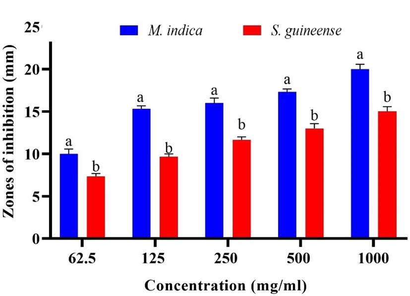
Antibacterial effects of the studied extracts were compared in the current study. However, at 31.25 mg/ml and 15.625 mg/ml concentration, S. guineense extracts did not show antibacterial activity. The antibacterial activity of M. indica ethanol extract showed significantly higher activity at all the tested concentrations than S. guineense extracts when tested against B. subtilis (p≤0.05; Figure 2).
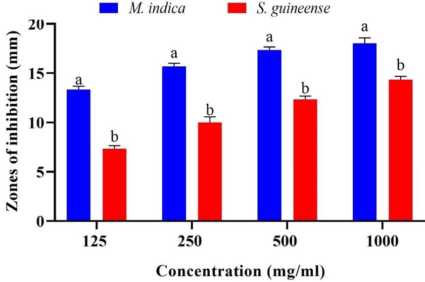
Similarly, M. indica ethanol extract showed significantly higher antibacterial effects at all tested concentrations compared to S. guineense when tested against S. aureus. The findings indicated that 125 mg/ml of ethanol extract of M. indica had significantly higher antibacterial activity against S. aureus compared with that of S. guineense (p≤0.05; Figure 3).
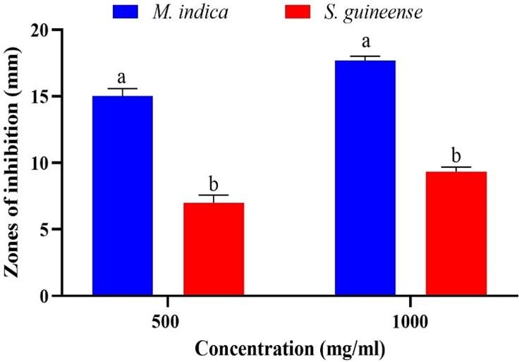
In comparison, Antibacterial activity exhibited by M. indica on S. typhi was significantly greater than that of S. guineense at concentrations of 1000, 500 and 250 mg/ml (p≤0.05; Figure 4). No comparisons were done for other tested doses of 125, 62.5, 31.25 and 15.625mg/ml since S. guineense extract did not show antibacterial activity.
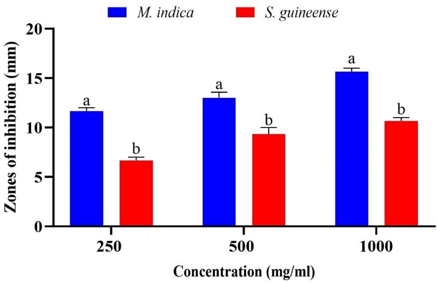
Similarly, Antibacterial activity exhibited by M. indica against E. coli was significantly greater than that of S. guineense at concentrations of 500 and 1000mg/ml (p≤0.05; Figure 5). There was no comparison done for other tested concentrations of 250, 125, 62.5, 31.25 and 15.625mg/ml since S. guineense extract did not show antibacterial activity.
Minimum inhibitory concentrations of ethanol extracts
MIC scores recorded for S. guineense extract against S. aureus and B. subtilis were statistically similar (p>0.05) but significantly lower than those against S. typhi and E. coli (p≤0.05). MIC scores for effects of M. indica extract on B. subtilis and S. aureus were relatively low but significantly distinct (p≤0.05). Further, MIC scores for M. indica effects on S. typhi and E. coli were statistically alike (p>0.05). However, they were significantly greater (p≤0.05) than those against B. subtilis and S. aureus (Table 1).
Table 1. Minimum inhibitory concentrations of ethanol extracts of M. indica and S. guineense.
Minimum bactericidal concentrations of ethanol extracts
M. indica extract demonstrated comparable (p>0.05) MBCs against E. coli and S. typhi but displayed significantly higher MBCs on B. subtilis and S. aureus (p≤0.05). M. indica achieved high MBC on S. typhi and E. coli but low MBCs on S. aureus and B. subtilis (Table 2). MBC scores exhibited by S. guineense extract on E. coli and S. typhi were comparable, as were MBC against S. aureus and B. subtilis (p≤0.05; Table 2).
Table 2. Minimum bactericidal concentrations of M. indica and S. guineense.
Qualitative phytochemical analysis of ethanolic extracts
Qualitative analysis of phytochemical constituents of M. indica ethanol extract showed the presence of saponins, quinones, tannins, flavonoids, alkaloids, steroids, tepenoids and phenolics while S. guineense extract contained saponins, quinones, flavonoids, alkaloids, glycosides, phenolics, steroids, terpenoids and tannins (Table 3). Fixed oils were absent in all studied extracts, and glycosides were not found in M. indica extract (Table 3).
Table 3. Qualitative phytochemical analysis of ethanolic extract of M. indica and S. guineense.
DISCUSSION
In the current study, ethanol extracts of M. indica and S. guineeense exhibited antibacterial effects. The ethanol extracts exhibited concentration-dependent inhibitory effects against the tested bacteria. This means that at higher concentrations, the extracts potently inhibited antibacterial activities. The ethanol extracts of M. indica and S. guineense exhibited significantly higher antibacterial effects against Gram positive bacteria (S. aureus and B. subtilis) than Gram negative bacteria (E. coli and S. typhi). This may be attributed to Gram negative bacteria possessing an extra lipopolysaccharide rich outer membrane, which gives them resistance to lipophilic compounds. In contrast, Gram positive bacteria do not have a resistant outer membrane but an outer peptidoglycan layer, which is a weak permeable barrier [17].
Findings of the present study corroborate with a previous research done on O. basilicum, U. dioica, T. serphyllium, and A. millefolium plants extracts on Gram positive bacteria, including B. cereus and S. aureus as well as Gram negative bacteria like S. typhi, E. coli and Pseudomonas aeruginosa where the extracts displayed more antibiotic efficacy on Gram positive bacteria compared to Gram negative bacteria [29]. In the same way, a study on the effects of methanol extract of Crocus sativus against S. aureus, Bacillus cereus, P. aeruginosa and E. coli where the extract had the highest antibacterial effect against S. aureus and C. sativus extract had the least antibacterial effect against P. aeruginosa [30].
M. indica seed extract inhibited the proliferation of Gram positive as well as Gram negative bacteria, which included B. subtilis, E. coli, S. aureus and S. typhi. These findings concur with those of a previous study done on M. indica leaf extract, which inhibited the proliferation of E. coli, S. pyogenase, P. mirabilis, S. aureus, S. pneumoniae, B. cereus, S. typhi, S. flexnerri and P. aeruginosa [31]. M. indica stem bark extract has shown other biological activities, including antiviral and anti-inflammatory efficacies [10]. Hannan et al. [32] also observed substantial inhibitory efficacy of M. indica leaf extract against S. typhi. In the current study, M. indica seed extract has proven efficacious against the tested bacteria with the greatest inhibitory effect against Gram positive bacteria [31].
Ethanol extract of S. guineense subdued proliferation of S. aureus and B. subtilis and slightly inhibited growth of Gram negative bacteria; S. typhi and E. coli. S. guineense extract subdued the growth of Gram-negative bacteria only at higher concentrations. This study is in line with a previous study on different Syzygium genus species, which are S. francissi, S. moorei, S. forte, S. wilsonii and S. puberulum methanol leaves extracts against E. coli, S. epidermidis, S. sonnei, S. aureus, and S. pyogenes. All extracts had an antibacterial effect on all bacteria strains, although Gram positive bacteria were slightly more susceptible [33].
MIC and MBC of the extracts were determined to further test the antibacterial effectiveness of the studied extracts. The M. indica seeds extract showed lower MIC values than S. guineense stem bark extracts. This suggests that M. indica seeds extract had higher antibacterial efficacy compared to S. guineense extracts [31]. The findings are consistent with a study on Ochroma pyramidale, Banisteriopsis caapii, Croton lechleri, Eugenia obtusifolia and Miconia salicifolia extracts against E. coli and S. aureus where low MIC scores were reported, which indicated a high antibacterial efficacy [34].
The MBC values obtained from ethanol extracts of M. indica and S. guineense against studied bacteria were greater than MIC values, indicating that the plant extracts exhibited bacteriostatic activity at higher dilutions and are bactericidal at lower dilutions. The findings corroborate with the study on phenolic compounds against S. pyogenes, where MBC values were higher than MIC values, indicating the presence of bactericidal compounds at higher concentrations [35].
These studied extracts are therefore suggested to be used as antibacterial agents and work by suppressing bacterial growth without necessarily killing them. They were also more effective on Gram positive bacteria than Gram negative bacteria. This study offers scientific motive for traditional uses of the four studied medicinal plants for the management of bacterial infections, especially those caused by Gram positive bacteria.
The antibacterial activities in this study of ethanol extracts of M. indica and S. guineeens could be associated with their constituent secondary metabolites. The secondary metabolites found in these studied extracts have been reported as growth inhibitors of both Gram positive bacteria as well as Gram negative bacteria [36, 37]. Gallic acid, quercetin, caffeic acid, coumarin, and catechol are phenolic compounds that possess antibacterial properties on Gram negative bacteria including P. aeruginosa and E. coli [38]. Quercetin inhibit the proliferation of S. aureus and E. coli [39].
Flavonoids have also been recorded to have antibacterial efficacy against V. cholerae, Shigella, S. mutans and other bacteria. Catechins have been demonstrated to inactivate cholera toxin produced by V. cholera [40]. Sophoraflavanone G and naringenin possess antibacterial efficacy against S. aureus and streptococci [41]. Alkaloids purified from Eclipta alba leaf are reported to have a significant antibacterial effect on Shigella boydii, P. aeruginosa, E. coli, S. aureus, and S. faecalis [42]. Saponins fractionated from Chenopodium quinoa have been documented to have antifungal and anti-inflammatory properties [43]. Tannin extracts have been reported as inhibitors of the growth of S. aureus, B. subitilis and Shigella dysenteriae [44]. Terpenoids have also been recorded to possess significant higher antibacterial and antifungal activities [45].
CONCLUSION
The ethanol extracts of S. guineense and M. indica showed in vitro antibacterial activity against E. coli, S. typhi, S. aureus and B. subtilis. The ethanol extracts of S. guineense and M. indica contain important phytochemical compounds associated with antibacterial activities. The ethanol extracts of S. guineense and M. indica could be utilized for the development of alternative remedies to treat bacterial infections. The study's findings led to the following suggestions: Bioassay-guided fractionation and isolation of bioactive antibacterial chemicals should be carried out, as well as evaluation of antibacterial activities on other harmful bacteria.
ACKNOWLEDGEMENT
Special gratitude to East African Community Scholarship Program funded by KFW and implemented by Inter-University Council for East Africa (IUCEA) and Adroit Consult International for funding this study.
AUTHOR CONTRIBUTIONS
This study was successful under the supervision of George Omwenga and Mathew Ngugi and technical assistance from James Kimani, Ibrahim Waweru and Paul Nyalo.
CONFLICTS OF INTEREST
There is no conflict of interest among the authors.
References
- [1]Aslam MS and Ahmad MS. Worldwide importance of medicinal plants: current and historical perspectives. Recent Adv. Biol. Med 2016; 02: 88
- [2]Hussein AR and El-Anssary AA. Plants secondary metabolites: the key drivers of the pharmacological actions of medicinal plants. Herb. Med., 2019.
- [3]Oladeji O. Natural Products : The characteristics and roles of medicinal plants : some important medicinal plants in Nigeria. An Indian Journal 2016; 12(3): 1–8.
- [4]Nahashon M. Conservation of wild-harvested medicinal plant species in Tanzania. 2013; 50.
- [5]Rajurkar N and Hande SM. Estimation of phytochemical content and antioxidant activity of some selected traditional indian medicinal plants. Indian J. Pharm. Sci 2011; 73(2): 146–151.
- [6]Ahmed D, Saeed R, Shakeel N, Fatima K, and Arshad A. Antimicrobial activities of methanolic extract of Carissa opaca roots and its fractions and compounds isolated from the most active ethyl acetate fraction. Asian Pac. J. Trop. Biomed 2015; 5(7): 541–545.
- [7]Okomo U, Akpalu EN, Le Doare K, Roca A, Cousens S, Jarde A, et al. Aetiology of invasive bacterial infection and antimicrobial resistance in neonates in sub-saharan Africa: A systematic review and meta-analysis in line with the strobe-ni reporting guidelines. Lancet Infect. Dis 2019; 19(11): 1219–1234.
- [8]Ebimieowei E and Ibemologi A. Antibiotics: classification and mechanisms of action with emphasis on molecular perspectives. Int. J. Appl. Microbiol. Biotechnol. Res 2016; 4: 90–101.
- [9]Salinitro M, Vicentini R, Bonomi C and Tassoni A. Traditional knowledge on wild and cultivated plants in the kilombero valley (Morogoro Region, Tanzania). J. Ethnobiol. Ethnomed 2017; 13(1): 1–14.
- [10]Singh R, Singh SK, Maharia RS, and Garg AN. Identification of new phytoconstituents and antimicrobial activity in stem bark of Mangifera indica (L.). J. Pharm. Biomed. Anal 2015; 105: 150–155.
- [11]Amri E and Kisangau DP. Ethnomedicinal study of plants used in villages around kimboza forest reserve in Morogoro, Tanzania. J. Ethnobiol. Ethnomed 2012; 8(1).
- [12]Djoukeng JD, Abou-Mansour E, Tabacchi R, Tapondjou AL, Bouda H, and Lontsi D. Antibacterial triterpenes from syzygium guineense (Myrtaceae). J. Ethnopharmacol 2005; 101(1-3): 283–286.
- [13]Chirchir DK, Cheplogoi PK, and Omolo JK. Chemical characterization of syzygium guineense ( myrtaceae ) stem bark extracts.2019; 8(3): 278–282.
- [14]Runyoro DK, Ngassapa OD, Matee MI, Joseph CC, and Moshi MJ. Medicinal plants used by tanzanian traditional healers in the management of candida infections. J. Ethnopharmacol 2006; 106 (2): 158–165.
- [15]Nikolic M, Vasic S, Djurdjevic J, Stefanovic O, and Comic L. Antibacterial and anti-biofilm activity of ginger 349 www.bsmiab.org/jabet Mavanza et al., J Adv Biotechnol Exp Ther. 2023 May; 6(2): 337-349 (Zingiber officinale (roscoe)) ethanolic extract. Kragujev. J. Sci 2014; 36 (36):129–136.
- [16]Hudzicki J. Kirby-Bauer disk diffusion susceptibility test protocol author information. Am. Soc. Microbiol 2016;1–13.
- [17]Debalke D, Birhan M, Kinubeh A and Yayeh M. Assessments of antibacterial effects of aqueous-ethanolic extracts of sida rhombifolia’s aerial part. Sci. World J 2018.
- [18]Grare M, Fontanay S, Cornil C, Finance C, and Duval RE. Tetrazolium salts for MIC determination in microplates: why? Which salt to select? How? J. Microbiol. Methods 2008; 75 (1): 156–159
- [19]Natta L, Orapin K, Krittika N and Pantip B. Essential oil from five zingiberaceae for anti food-borne bacteria. Int. Food Res. J 2008; 15(3): 337–346
- [20]Bandiola TM. Screening of Medicinal Plants. Int. J. Pharm 2018; 8(1): 137–143
- [21]Abera B, Adane L, and Mamo F. Phytochemical investigation the root extract of syzygium guineense and Isolation of 2 , 3 , 23- trihydroxy methyl oleanate. J. Pharmacogn. Phyochemistry 2018; 7(2): 3104–3111
- [22]RY and Agarwala M. Phytochemical analysis of some medicinal plants. Phytology 2011; 3(8): 1–5.
- [23]Rajesh KD, Vasantha S, Rajesh NV and Panneerselvam A. Qualitative and quantitative phytochemical analysis in four pteridophytes. Int. J. Pharm. Sci. Rev. Res 2014; 27(2): 408–412.
- [24]Soni A and Sosa S. Phytochemical analysis and free radical scavenging potential of herbal and medicinal plant extracts. J. Pharmacogn. Phytochem. JPP 2013; 22(24): 22–29.
- [25]Santhi K and Sengottuvel R. Qualitative and quantitative phytochemical analysis of moringa concanensis nimmo. Int. J. Curr. Microbiol. Appl. Sci 2016; 5(1): 633–640.
- [26]Medeo N, Haque M, Sahira K and Cathrine L. General techniques involved in phytochemical analysis related papers extraction, isolation and characterization of bioactive compounds from plant. Int. J. Adv. Res. Chem. Sci 2015; 2 (4): 25–32.
- [27]Hanafi H, Irawan C, Rochaeni H, Sulistiawaty L, Roziafanto AN and Supriyono. Phytochemical screening, lcms studies and antidiabetic potential of methanol extracts of seed shells of archidendron bubalinum (jack) i.c. nielson (julang jaling) from Lampung, Indonesia. Pharmacogn. J 2018; 10(6): S77–S82.
- [28]Thaipong K, Boonprakob U, Crosby K, Cisneros-Zevallos L and Hawkins DB. Comparison of ABTS, DPPH, FRAP, and ORAC assays for estimating antioxidant activity from guava fruit extracts. J. Food Compos. Anal 2006; 19(6-7): 669–675.
- [29]Bobis O, Dezmirean DS, Tomos L, Chirila F and Al. Marghitas L. Influence of phytochemical profile on antibacterial activity of different medicinal plants against gram-positive and gram-negative bacteria. Appl. Biochem. Microbiol 2015; 51(1): 113–118.
- [30]Jafari-sales A and Pashazadeh M. Antibacterial effect of methanolic extract of saffron petal (Crocus sativus L.) on some standard gram positive and gram negative pathogenic bacteria in vitro. Curr. Perspect. Med. Aromat. Plants 2020; 3(1): 1–7.
- [31]Doughari JH and Manzara S. In vitro Antibacterial activity of crude leaf extracts of mangifera indica linn. African J. Microbiol. Res 2008; 2(4): 67–72.
- [32]Hannan A, Asghar S, Naeem T, Ullah MI, Ahmed I, Aneela S, et al. Antibacterial effect of mango (mangifera indica linn.) leaf extract against antibiotic sensitive and multi-drug resistant salmonella typhi. Pak. J. Pharm. Sci 2013; 26(4): 715–719.
- [33]Chikowe G, La M and Cock B. Antibacterial activity of syzygium species. pharmacogn. Commun 2013; 3(4): 365–367.
- [34]Bussmann R. W, Grenn A, Sharon D, Chait G, Diaz D, Pourmand K, et al. Minimum inhibitory concentration of medicinal plants used in northern peru as antibacterial remedies. J. Ethnopharmacol 2010; 132(1): 101–108.
- [35]Macé S, Truelstrup L, and Rupasinghe HP. Anti-bacterial activity of phenolic compounds against Streptococcus pyogenes. Medicines 2017; 4(2): 25.
- [36]Saxena M, Saxena J, Nema R, Singh D and Gupta A. Phytochemistry of medicinal plants. Med. Plants Cent. Asia Uzb. Kyrg 2013; 1(6): 13–14.
- [37]Pandey S. Antibacterial and Antifungal Activities of Ocimum gratissimum L. Int. J. Pharm. Pharm. Sci 2017; 9(12): 26; doi: 10.22159/ijpps.2017v9i12.22678.
- [38]Tyagi B, Dubey A, Verma AK, and Tiwari S. Antibacterial activity of phenolics compounds against pathogenic bacteria. Int. J. Pharm. Sci. Rev. Res 2015; 35(1): 16–18.
- [39]Lin JK and Weng MS. Flavonoids as nutraceuticals. Sci. Flavonoids 2006; 7: 213–238.
- [40]Karak P. Biological activities of flavonoids: An Overview. Int. J. Pharm. Sci. Res 2019; 10(4): 1567–1574.
- [41]Kumar S and Pandey AK. Chemistry and biological activities of flavonoids: an overview. Sci. World J 2013; 145–148.
- [42]Gurrapu S and Mamidala E. In vitro Antibacterial activity of alkaloids isolated from leaves of eclipta alba against human pathogenic bacteria. Pharmacogn. J 2017; 9(4): 573–577.
- [43]Kuljanabhagavad K and Wink M. Biological Activities and chemistry of saponins from chenopodium quinoa willd. Phytochem. Rev 2009; 8(2): 473–490.
- [44]Kurhekar JV. Tannins – antimicrobial chemical components. Int. J. Technol. Sci 2016; 6: 5–9.
- [45]Zhang L and Demain A. Natural product: drug discovery and therapeutic medicine. Spr Sci Med 2016; 3-29