Exposure to tobacco (Nicotiana tabacum) extract and cigarette smoke induces hematological and histopathological alterations in Swiss albino mice
Abstract
Tobacco smoking is a widely practiced recreational activity and is associated with numerous health risks. Hence, the present study was conducted on Swiss albino mice to assess the impact of an aqueous extract of tobacco and cigarette smoke on behavioral, hematological, and histopathological parameters of the lungs, liver, and kidney. The mice were divided into control, Tobacco nicotiana (TN), and cigarette smoke (CS) groups (n=6). The TN group received aqueous tobacco orally at a dose of 5 ml/kg BW for 21 days, whereas the CS group was exposed to inhalational commercial cigarette smoke for the same duration. On day 22, blood samples and visceral organs were collected to evaluate hematological and histopathological parameters in euthanized mice. The total erythrocyte count was significantly elevated (P<0.05) in both groups. In contrast to the control group, the total leukocyte count was lower in the CS group (P>0.05) than in the TN group, but the hemoglobin showed no significant changes. Lower body weight, aggressive behavior, and weakness were observed in the treated groups. Histopathologically, the lung revealed emphysematous changes, alveolar septal thickening, inflammatory cell infiltration, congestion, and thrombus formation in pulmonary artery. The liver exhibited hepatocyte degeneration with karyomegaly and karyorrhexis, derangement of the hepatic cords and sinusoids, and central vein congestion. Derangement of renal tubules, edema, and glomerular degeneration were observed in the kidney. These observations lead us to postulate that tobacco and cigarette smoke exert detrimental effects on the structure of some vital organs in the body after direct or indirect uptake.
INTRODUCTION
Tobacco smoking is a widely prevalent form of recreational drug being utilized either directly or indirectly by over one billion people globally, of whom the majority live in developing countries. The toxic nature of all tobacco products poses significant risks to human health, with no safe level of exposure established. Cigarette smoking, the most common method of tobacco consumption worldwide, contributes to a major global public health crisis, causing over 8 million deaths annually, equivalent to approximately one death every 6 seconds [1]. Additionally, secondhand smoking causes 1.2 million deaths each year [2].
The World Health Organization's Framework Convention on Tobacco Control, 2009, defines tobacco products as those entirely or partially derived from the tobacco leaf, intended for activities such as smoking, sucking, chewing, or snuffing [3]. Among those, smoking is the most convenient method of tobacco consumption [4], with nicotine being the most prominent and highly addictive phytochemical found in tobacco [5]. In the case of cigarette smoking, the inhalation of heated aerosols containing active nicotine alkaloids allows their absorption into the small airways and alveoli of the lungs and subsequent increase in blood concentration within 10 seconds, affecting vital organs such as the heart, lungs, brain, and others that increase the risk of various diseases [6]. Smoking also impacts individuals' overall well-being, financial status, and those near to smokers. On average, a single cigarette contains approximately 2 mg of absorbable nicotine [7]. Moreover, one gram of tobacco contains 4.4 to 25 milligrams of nicotine [8]. The calculated lower limit for nicotine consumption in adults is estimated to be 6.5–13 mg/kg [9]. However, high doses of nicotine can lead to poisoning, organ failure, and respiratory muscle paralysis, with a lethal dosage of approximately 127 mg/kg in mice [10]. Nicotine from tobacco can be acquired through gas, gum, and aqueous forms, with chewing gum delivering lower nicotine compared to cigarettes [11]. The particulate phase of tobacco smoke contains carcinogens, ganglion stimulators and depressors, tumor accelerators, and irritants, while the gas phase comprises substances impairing oxygen transport and utilization, as well as carcinogens and irritants [12].
The development of tobacco-related diseases is influenced by both exposure time and dosage [13]. The detrimental effects of tobacco smoking on health can be categorized into three main areas: lung diseases (e.g., chronic obstructive pulmonary disease [COPD], pulmonary fibrosis, bronchitis, emphysema, and asthma) [14], malignancies (specifically lung, oral, esophageal, stomach, pancreatic, colorectal, and bladder cancers) [15], and cardiovascular diseases (including myocardial infarctions and cerebrovascular accidents) [16]. Tobacco smoking also increases the risk of liver fibrosis and liver cancer [17], as well as causes adverse effects on renal function, leading to chronic kidney disease and renal cancer [18]. A single cigarette contains approximately 4,000 chemicals, and upon inhalation, these substances generate reactive oxygen species (ROS) and oxidative stress in lung tissue, resulting in emphysematous changes [19]. The hepatotoxicity caused by cigarette chemicals induces oxidative stress in liver tissue, activating stellate cells and leading to fibrosis [20]. The potential mechanism of kidney damage associated with smoking involves increased blood pressure, alterations in intra-renal hemodynamics, and activation of the renin-angiotensin system [21, 22] .
Despite the established health risks associated with tobacco smoke exposure in humans, limited research has examined the effects of tobacco use in aqueous form on various vital organs. Therefore, our study aims to investigate the detrimental effects of continuous exposure to tobacco in aqueous and smoking forms on the lungs, liver, and kidney in Swiss albino mice. We hypothesize that both forms of tobacco exposure exhibit harmful effects on these organs.
MATERIALS AND METHODS
Ethical approval
This study was accomplished according to the institutional rules and regulations for animal research, care, and use. The experimental procedure was approved by the animal research ethical committee of the Faculty of Veterinary Medicine and Animal Science (Reference no. FVMAS/ AREC/0028/2023).
Study location
The study was conducted in collaboration with the laboratory of Anatomy & Histology and Pathobiology of the faculty of Veterinary Medicine and Animal Science in Bangabandhu Sheikh Mujibur Rahman Agricultural University, Bangladesh.
Animals
The experimental male Swiss albino mice were procured from the International Center for Diarrheal Disease Research (icddr’b), Mohakhali, Dhaka, Bangladesh. According to the registered veterinarian from the icddr'b, all the mice were in good condition and free of any outward defects. After procurement, all the mice were kept under close observation to acclimate to the new environment for a period of one week prior to the commencement of the experiment. The designed Swiss albino mice were maintained on a 12:12-h light and dark cycle with controlled temperature (70–740 °F) and humidity (45–60%) at the animal facility of the faculty of veterinary medicine and animal science, with access to water and mouse pellets ad libitum.
Preparation of an aqueous extract of tobacco
Tobacco and cigarettes were purchased from the local market in Bangladesh. Upon procurement, the tobacco was air-dried at room temperature and subsequently ground into a fine powder using a mortar and pestle. The fine powder was then sieved through a mesh to eliminate any coarse particles. For the preparation of the tobacco solution, 5g of the fine powder was mixed with 50 mL of distilled water and stirred using an electric shaker at room temperature (25°C) for 10 minutes. The resulting mixture was filtered using Whatman filter paper and stored in a refrigerator at 4°C for future use. The dosage of the tobacco solution was determined based on the body weight of the mice.
Preparation of cigarette smoke
As per the manufacturer's specifications, each cigarette contains 1 mg of nicotine. To facilitate the exposure of cigarette smoke, we constructed compact glass chambers (dimensions: 45×30×30 cm) equipped with controlled negative airflow ventilation. Additionally, control groups for cigarette smoke were exposed to ambient fresh air in a controlled chamber.
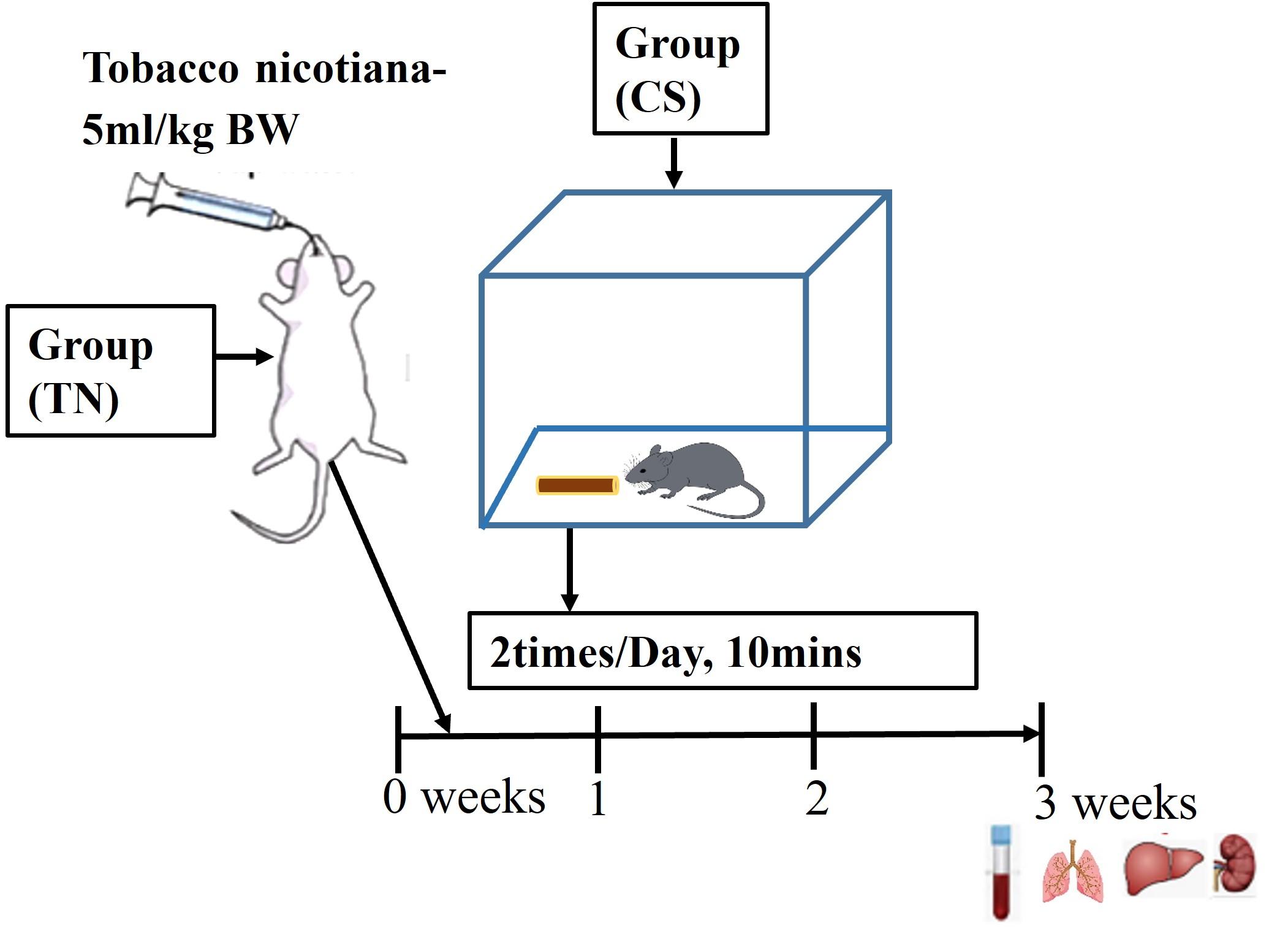
Experimental approach
A total of 18 adult male Swiss albino mice, aged 5 weeks and weighing between 27 and 33 g, were selected as the animal model for this study. The initial body weights of all mice were recorded prior to the initiation of the experiment. Throughout the study, the body weight of each mouse was monitored weekly to ensure accurate adjustment of the tobacco dosage. Body weight was evaluated on weeks 0 (days 0), 1, 2, and 3. The mice were divided into three (3) experimental groups, with six mice in each group (n = 6).
The control (Cont) group received normal drinking water orally via syringe and was exposed to fresh environmental air continuously for a duration of 21 days. The tobacco nicotiana group (TN) received an aqueous extract of tobacco at a dose of 5 ml/kg BW orally once daily via syringe. This treatment was carried out for a period of 21 days. Prior to exposure to cigarette smoke, all mice in the cigarette smoke group (CS) were housed in glass chambers. They were then subjected to the inhalation of smoke from six commercial cigarettes, twice daily (at 9:00 AM and 4:00 PM), for 10 minutes each session. This exposure regimen was maintained for a duration of 21 days (Figure 1).
Observation of behavioral changes
During the experiment, mice were closely monitored and watched to look for any irregularities in their behavior. Body weight was measured using a digital balance, and eye inspection was employed to track the mice's behavior towards one another, their intake of food and water, and the condition of their coats. Following tobacco intake and cigarette smoke, the nature and onset of effects, as well as any deaths were meticulously documented by following a formula.
% = ![]() ×100
×100
Collection of samples, hematological and morphological analyses
Following the final exposure to aqueous tobacco and cigarette smoke, each animal was euthanized under the effects of xylazine and ketamine at a dose rate of 15 mg/kg xylazine and 100 mg/kg ketamine intraperitoneally via commercial injection. Subsequently, 2 mL of blood was collected from the cardiac puncture using a 5 mL disposable syringe. The blood samples were used for the estimation of various blood chemical parameters, including total erythrocyte count (TEC), total leukocyte count (TLC), and hemoglobin percentage (Hb%). The number of red blood cells (RBC) and white blood cells (WBC) were calculated based on the counted cells, where the RBC count was multiplied by 1000 and expressed in millions per cubic mm, and the WBC count was multiplied by 50 and expressed in thousands per cubic mm. Total erythrocyte count (TEC), total leukocyte count (TLC), and hemoglobin percentage (Hb%) were determined as per methods cited by Coffin (1955) [23].
Upon sacrificing each animal, the lungs, liver, and kidney were meticulously dissected for histopathological examination. Following the collection of tissue samples measuring 5 mm2 from the appropriate organs, the samples were preserved in a 10% neutral buffer formalin (NBF) for 48 hours. After that, the samples were washed in 10% phosphate buffer solution for 3 hours and subjected to dehydration using a series of ascending alcohol grades (70%, 80%, 90%, 95%, 100% for two hours each, and finally 100% overnight). Following dehydration, the samples were cleared by xylene and embedded in paraffin. The paraffin-embedded blocks were cut into 4 µm thick sections using a rotary microtome. These sections were subsequently stained with Meyer's hematoxylin and eosin (H&E). To protect the sections, a thin cover slip was attached to each slide using a mounting medium called 'DPX'. Histopathological observations were made using a Leica DM500 binocular microscope. All images were transferred to Adobe Photoshop Elements 15 (Adobe Systems, CA, USA) where adjustments were made solely to brightness and contrast.
Statistical analysis
Statistical analyses were conducted using Microsoft Excel (Microsoft Corp., Washington, USA). Descriptive data are presented as the mean ± SEM. A one-way ANOVA was employed for statistical assessment. Statistical significance was determined at a significance level of P≤0.05.
RESULTS
Effects of tobacco extract and cigarette smoke on behavior modifications
Throughout the entire experimental period, the mice in the control group exhibited normal health, vitality, and activity. In the TN group, mice exhibited aggressive behavior, apathy, and weakness. Conversely, the CS group, exposed to cigarette smoke, displayed signs of dissatisfaction, roughness of the hair coat, increased thirst, and weakness. No instances of mortality were observed (Table 1).
Table 1. Behavioral changes in experimental animals.
Effects of tobacco extract and cigarette smoke on body weight
The mean body weight of mice in the control group at the beginning and end of the experiment was 32.16±0.30 g and 42.08±0.80 g, respectively (Table 2). Total body weight was significantly (P<0.05) affected in the treated groups exposed to aqueous tobacco and cigarette smoke. However, at the end of the experiment, the body weights of TN and CS groups were measured accordingly, 40.33±0.42 g and 40.87±0.69 g, which were lower than the control group (Table 2).
Table 2. Effects of tobacco extract and cigarette smoke on body weight.
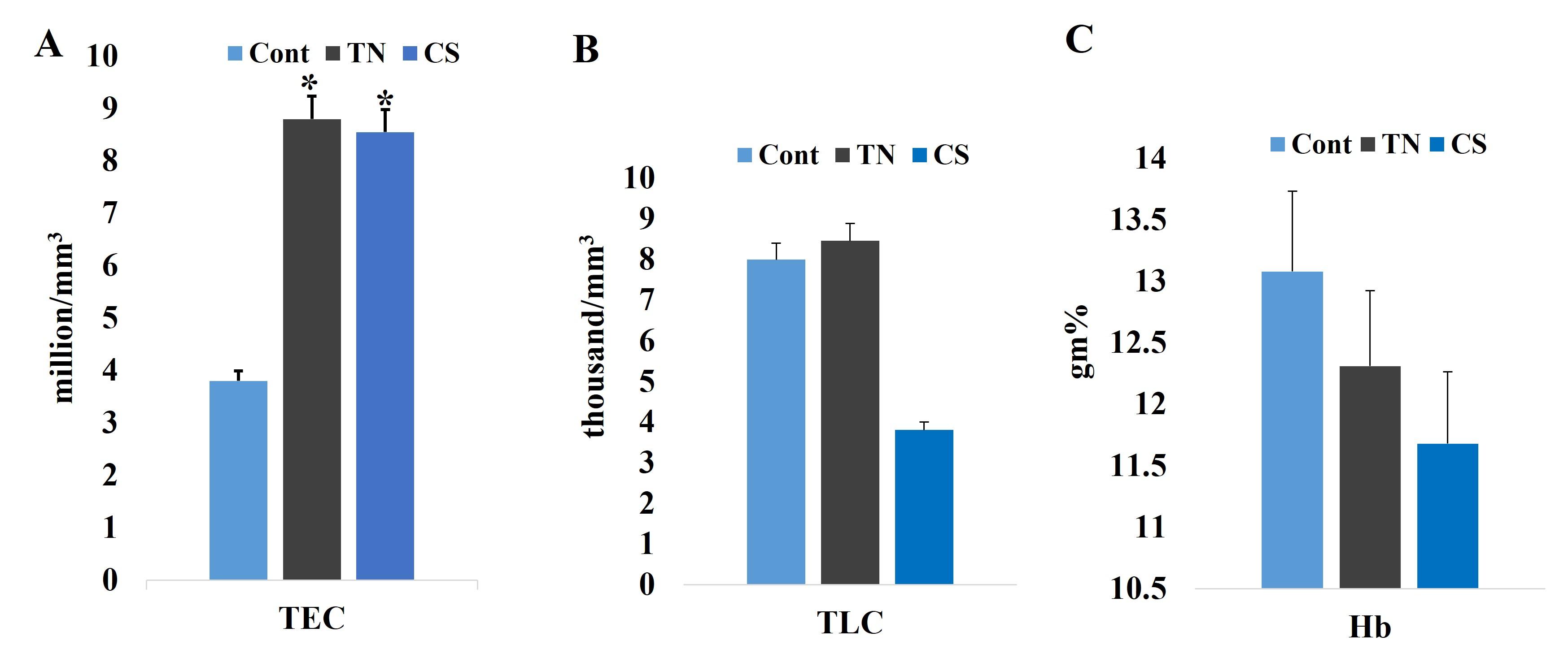
Effects of tobacco extract and cigarette smoke on hematological parameters
The hematological parameters (TEC, TLC, and Hb) were assessed after a 21-day treatment period. Following exposure to tobacco and cigarette smoke, the blood parameters, especially TEC value, revealed negative results. In the Control group, the mean TEC, TLC, and Hb% values were 3.80±0.09 million/mm3, 8.0±0.12 thousand/mm3, and 13.08±0.20 g/dL, respectively. However, exposure to tobacco and cigarette smoke significantly increased (P<0.05) TEC values in the TN and CS groups than the control group and the values were 8.80±0.45 million/ mm3 and 8.55±0.39 million/ mm3, respectively. However, the TLC value was lower in the CS group (3.81±0.27 thousand/ mm3) than to the TN group (9.46±2.13 thousand/ mm3). The mean Hb% values in the TN and CS groups were 12.31±0.55 g/dL and 11.68± 0.41 g/dL, which showed no significant changes compared to the control group (Figure 2).
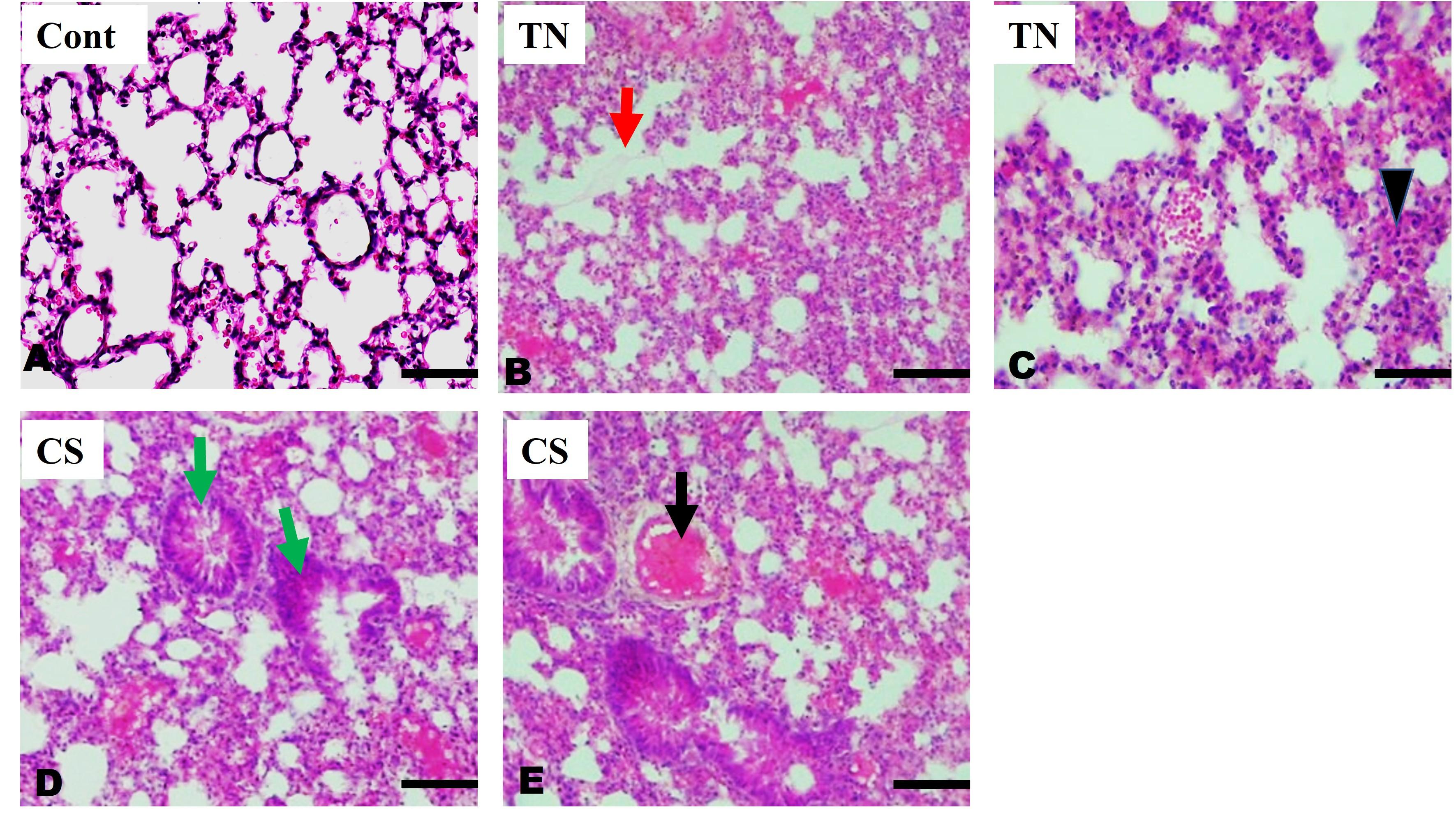
Effects of tobacco extract and cigarette smoke on lung parenchyma
The histology of lung tissues in the control group exhibited normal alveoli and alveolar ducts lined by pulmonary pneumocytes (Figure 3A). However, in the treated group, significant alterations were observed upon microscopic examination, including the disruption of pulmonary parenchymal architecture characterized by enlarged air spaces with the development of emphysematous changes, alveolar septal thickening, and infiltration of inflammatory cells in the TN group (Figure 3B-C). Additionally, distortion of bronchiolar smooth muscle with congestion, thrombus formation in the pulmonary artery, and thick alveolar septa were observed in the CS group (Figure 3D-E).
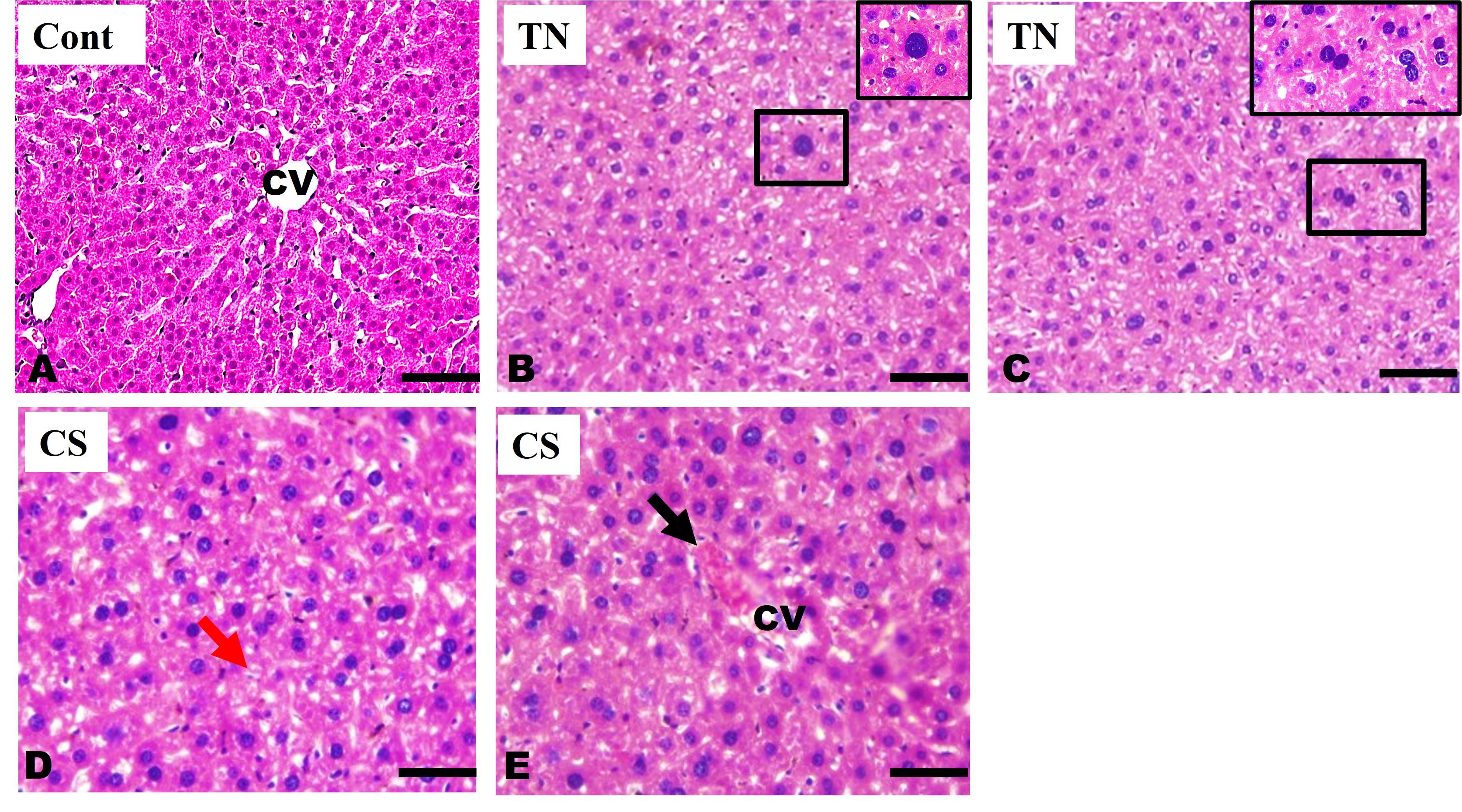
Effects of tobacco extract and cigarette smoke on liver parenchyma
The histological examination of the liver parenchyma in the control group revealed a normal hepatic lobule structure, characterized by a centrally located central vein and well-organized hepatic cords formed by radiating hepatocytes, along with intact sinusoids (Figure 4A). However, the treated groups exhibited certain alterations. In the TN group, microscopic observation revealed hepatocyte degeneration accompanied by an increase in nuclear size (karyomegaly) and nuclear karyorrhexis (Figure 4B-C). Conversely, the CS group exhibited disruption of hepatic cords and irregular sinusoidal structures and dilatation of the central vein with congestion (Figure 4D-E).
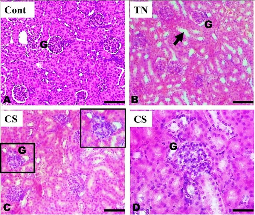
Effects of tobacco extract and cigarette smoke on kidney parenchyma
The histological analysis of the kidney parenchyma in the control group revealed well-organized renal corpuscles characterized by densely packed, rounded glomeruli. These glomeruli were surrounded by double-walled epithelial Bowman's capsules, and the proximal convoluted tubules and distal convoluted tubules exhibited a well-structured appearance (Figure 5A). In contrast, the TN group displayed a range of histological alterations, including the disruption of renal tubules and the presence of interstitial edema, which affected the separation of the tubules (Figure 5B). Whereas the CS group showed disorganization and degeneration of the glomeruli (Figure 5C), an increased number of swollen endothelial cells were observed, resulting in the occlusion of capillary lumens. As a result, the swollen capillary tufts completely obliterated Bowman's space (Figure 5D).
DISCUSSION
In this study, we observed noxious effects of aqueous extracts of tobacco and CS on behavioral, hematological, and pathological changes in the lungs, liver, and kidney of Swiss albino mice. Tobacco refers to the processed leaves of the Nicotiana tabaccum plant, which is widely cultivated and consumed worldwide through smoking, chewing, snuffing, or dipping [24]. CS is known to contain harmful chemicals that are toxic and carcinogenic, making it a significant risk factor for various diseases [25]. Exposure to tobacco and cigarette smoke is strongly linked to the development of several diseases in lungs, such as lung cancer, chronic obstructive pulmonary disease, and pulmonary fibrosis [26]. Additionally, tobacco smoking has negative impacts on liver and kidney function, leading to hepatotoxicity, hepatic fibrosis, hepatic cancer (hepatocellular carcinoma), renal cancer, and chronic kidney diseases [27, 12].
Smoking is a leading cause of morbidity and mortality worldwide, and it has detrimental effects on health [28]. Both smoking and being underweight have significant negative impacts on overall well-being. Tobacco consumption has been found to acutely increase energy expenditure, leading to a decrease in appetite [29]. In our study, body weight measurement was found to be lower among the tobacco and cigarette smoking groups. Previous studies have indicated that cigarette smokers tend to have a lower body weight compared to nonsmokers due to an increasing metabolic rate and decreasing caloric absorption [30]. Williamson et al. (1991) [31] reported that smokers have lower body weights, or body mass index (BMI; in kg/m2), than nonsmokers.
When people use smokeless tobacco, the main phytochemical nicotine remains in their blood longer than when they smoke. The level of nicotine remains in the blood depends on the amount of nicotine in the smokeless tobacco products and with their pH [32]. In our study, hematology results indicated a significant increase in TEC in the TN and CS groups compared to the control group (P≤0.05). However, TLC values were lower in CS group than to the TN group. Although the mean value of Hb% did not show any significant changes. According to previous study, aqueous extracts of Nicotiana tabacum increased the values of RBC, Hb, and hematocrit but did not significantly affect mean corpuscular volume, mean corpuscular hemoglobin, and mean corpuscular hemoglobin concentration compared to the control [33]. Although consumption of aqueous extract of tobacco decreased the value of RBC, hematocrite, and Hb than normal range in albino rats, it suggested the advancement of anemia [24].
Cigarette smoking leads to various diseases due to its high concentration of harmful and carcinogenic substances [34]. Specifically, it is directly linked to the development of fatal lung diseases. Our study revealed that exposure to tobacco and cigarette smoke resulted in damage to the alveoli of the lungs that led to emphysematous changes. Previous research has consistently highlighted cigarette smoke as a major risk factor for the development of emphysema, a key component of COPD. Cigarette smoke induces the destruction of alveolar cells and triggers oxidative stress and arylation processes, which are known to contribute to the development of emphysema [35]. Studies have also demonstrated that exposure to tobacco smoke for extended periods can lead to alveolar destruction, epithelial proliferation, increased alveolar macrophages, thickening of the vascular wall, and congestion [36]. Moreover, exposure to cigarette smoke and polyhexamethylene guanidine caused severe lung injury with inflammation and subsequent fibrosis [26].
Cigarette smoking also causes structural changes to the liver and kidneys. It induces the synthesis of ROS, leading to cellular damage in the liver [37]. In this study, liver showed degeneration of hepatocytes, disruption of hepatic cords, abnormal sinusoids, and central vein dilatation with congestion in treated groups. Hepatocytes are vital functional cells in the liver responsible for maintaining blood glucose levels and energy production. Damage to hepatocytes, as evidenced by increased nuclear size and nuclear fragmentation, affects liver function [13]. Similar studies have reported degeneration, apoptosis of hepatocytes, reduced sinusoidal lining cells, inflammation in the central vein, and vacuolation in the hepatic parenchyma due to tobacco smoking [13, 24]. Nicotine administration in mice provokes hepatocyte swelling, and perivascular infiltration of inflammatory cells [38]. Although we did not assess oxidative and inflammatory stress markers in this experiment, earlier studies have shown that the use of cigarette smoke leads to elevated pro-inflammatory and oxidative markers in the brain regions of rats [39].
The kidney is a vital organ responsible for regulating the body's water and salt balance and eliminating foreign substances from the blood. Our study showed exposure to tobacco and cigarette smoke caused derangement of renal tubules, interstitial tissue edema, glomerular degeneration, and obliterated Bowman's space in treated groups. Previous studies have shown that inhalation of cigarette smoke results in renal tissue necrosis and cortical vascular congestion [38, 40]. The proximal convoluted tubules showed severe cellular degradation, potentially affecting their structural integrity [13]. Smoking-induced renal damage occurs through acute hemodynamic changes such as increased blood pressure, intraglomerular pressure, and endothelial cell dysfunction [21]. Activation of the sympathetic nerve and the renin-angiotensin system also plays a potential role in renal damage due to smoking [22].
Chronic exposure to cigarette smoke induces systemic oxidative stress, resulting in the production of profibrotic factors. DNA damage markers were found to be significantly higher in the heart, liver, and lungs of rats exposed to cigarette smoke [41]. Although earlier studies have observed harmful effects of cigarette smoking in various organs of individuals, this study was performed to know the deleterious effects of consumption of aqueous tobacco and passive smoking in vital organs of the body. Further research is needed to investigate the different dose-dependent effects of aqueous tobacco. We used Swiss albino mice in this research because mice are considered as mimic animals with physiological similarities to humans.
CONCLUSION
In comparison to cigarette smoke, tobacco has some medicinal potential, but this study provides helpful information about the detrimental effects of aqueous tobacco and passive smoking in Swiss albino mice that showed a significant increase in red blood cell counts, weakened mice, and histological changes in the lungs, liver, and kidney. This research finding suggests that we should be cautious about the usage of tobacco, either in aqueous or smoking form.
ACKNOWLEDGEMENT
We acknowledge the Department of Anatomy and Histology as well as Department of Pathobiology, Faculty of Veterinary Medicine and Animal Science, Bangabandhu Sheikh Mujibur Rahman Agricultural University, Gazipur-1706, Bangladesh for laboratory support.
AUTHOR CONTRIBUTIONS
NJ: Conceptualization, data curation, formal analysis, investigation, methodology, software, visualization, writing - original draft, writing - review & editing. SA and MTH: Formal analysis, investigation, methodology. MAAM: Formal analysis, investigation, visualization, writing - review& editing. All authors approved the final version of the manuscript.
CONFLICTS OF INTEREST
There is no conflict of interest among the authors.
References
- [1]WHO (2019). WHO report on the global tobacco epidemic: Offer help to quit tobacco use.
- [2]World Population Review (2023). Smoking Rates by Country 2023. worldpopulationreview.com.
- [3]WHO (2009). History of the World Health Organization Framework Convention on Tobacco Control.
- [4]Wigand JS. ADDITIVES , CIGARETTE DESIGN and TOBACCO PRODUCT REGULATION. Tobacco product regulation group. 2006.
- [5]International Agency for Research on Cancer (2007). Monographs on the Evaluation of Carcinogenic Risks to Humans - Volume 89. Smokeless Tobacco and Some Tobacco-specific N-Nitrosamines.
- [6]Benowitz NL, Hukkanen J, et al. Nicotine chemistry, metabolism, kinetics and biomarkers. Handb Exp Pharmacol. 2009; 192: 29-60.
- [7]Mayer B. How much nicotine kills a human? Tracing back the generally accepted lethal dose to dubious self-experiments in the nineteenth century. Archives of Toxicology. 2014; 88: 5–7.
- [8]Richter P, Hodge K, et al. Surveillance of moist snuff: total nicotine, moisture, pH, un-ionized nicotine, and tobacco-specific nitrosamines. Nicotine & Tobacco Research. 2008; 10(11): 1645–1652.
- [9]Royal College of Physicians. Nicotine without smoke: tobacco harm reduction. Pediatrics. 2016.
- [10]Schraufnagel DE. Electronic Cigarettes: Vulnerability of Youth. Pediatric Allergy, Immunology, and Pulmonology. 2015; 28(1): 2–6.
- [11]West RJ, Jarvis M, et al. Effect of nicotine replacement on the cigarette withdrawal syndrome. British Journal of Addiction. 1984; 79(2): 215-219.
- [12]El-Zayadi AR. Heavy smoking and liver. World Journal of Gastroenterology. 2006; 12(38): 6098-6101.
- [13]Adedayo AD, Musa A, et al. Histological study of smoke extract of Tobacco nicotiana on the heart, liver, lungs, kidney, and testes of male Sprague-Dawley rats. Nigerian Medical Journal. 2011; 52(4): 217-222.
- [14]Patel RR, Ryu JH, et al. Cigarette Smoking and Diffuse Lung Disease. Drugs. 2008; 68: 1511–1527.
- [15]Sasco AJ, Secretan MB, et al. Tobacco smoking and cancer: a brief review of recent epidemiological evidence. Lung Cancer. 2004; 45: S3–S9.
- [16]Wilson PWF. Smoking, smoking cessation, and risk of cardiovascular disease. Current Treatment Options in Cardiovascular Medicine. 2006; 8: 276–281.
- [17]Rutledge SM, Asgharpour A. Smoking and liver disease. Gastroenterology & Hepatology. 2020; 16(12): 617-625.
- [18]Jaimes EA, Tian R.-X, et al. Nicotine Augments Glomerular Injury in a Rat Model of Acute Nephritis. American Journal of Nephrology. 2009; 29(4): 319–326.
- [19]Barnes PJ, Shapiro SD, et al. Chronic obstructive pulmonary disease: molecular and cellular mechanisms. European Respiratory Journal. 2003; 22: 672–688.
- [20]Husain K, Scott BR, et al. Chronic ethanol and nicotine interaction on rat tissue antioxidant defense system. Alcohol. 2001; 25(2): 89-97.
- [21]Orth SR. Effects of Smoking on Systemic and Intrarenal Hemodynamics. Journal of the American Society of Nephrology. 2004; 15: S58–S63.
- [22]Orth SR. Smoking – A Renal Risk Factor. Nephron. 2000; 86: 12-26.
- [23]Coffin DI. Manual Veterinary Clinical Pathology 3rd edition. Comstock Publishing Associates. Inc. Newwork, 1955.
- [24]Shekins O, Dorathy E, et al. Phytochemical Screening of Tobacco (Nicotiana tabacum) and Its Effects on Some Haematological Parameters and Histopathology of Liver and Brain in Male Rats. International Journal of Biochemistry Research & Review. 2016; 14(4): 1–9.
- [25]Eldridge A, Betson TR, et al. Variation in tobacco and mainstream smoke toxicant yields from selected commercial cigarette products. Regulatory Toxicology and Pharmacology. 2015; 71: 409–427.
- [26]Shin Y-J, Kim S-H, et al. Exposure to cigarette smoke exacerbates polyhexamethylene guanidine-induced lung fibrosis in mice. The Journal of Toxicological Sciences. 2021; 46(10): 487–497.
- [27]Black HR, Zeevi GR, et al. Effect of Heavy Cigarette Smoking on Renal and Myocardial Arterioles. Nephron. 1983; 34: 173–179.
- [28]Mokdad AH. Actual Causes of Death in the United States, 2000. JAMA Network. 2004; 291(10): 1238-1245.
- [29]Hofstetter A, Schutz Y, et al. Increased 24-Hour Energy Expenditure in Cigarette Smokers. The New England Journal of Medicine. 1986; 314: 79–82.
- [30]Chiolero A, Wietlisbach V, et al. Clustering of risk behaviors with cigarette consumption: A population-based survey. Preventive Medicine. 2006; 42(5): 348–353.
- [31]Williamson DF, Madans J, et al. Smoking cessation and severity of weight gain in a national cohort. The New England Journal of Medicine. 1991; 324(11): 739-745.
- [32]Omotoso GO, Kadir RE, et al. Exposure to cigarette smoke altered the cytoarchitecture and antioxidant activity of the frontal cortex in wistar rats. International Journal of Biological and Chemical Sciences. 2013; 7(4): 1595-1601.
- [33]Fafioye OO. Acute and sub-acute toxicities of five plant extracts on white tilapia, Oreochromis niloticus (Trewavas). International Research Journal of Agricultural Science and Soil Science. 2012; 2(13): 525–530.
- [34]Eldridge A, Betson TR, et al.Variation in tobacco and mainstream smoke toxicant yields from selected commercial cigarette products. Regulatory Toxicology and Pharmacology. 2015; 71: 409–427.
- [35]Ghosh A, Ganguly S, et al. Causation of Cigarette Smoke–Induced Emphysema by p-Benzoquinone and Its Prevention by Vitamin C. American Journal of Respiratory Cell and Molecular Biology. 2015; 52(3): 315–322.
- [36]Dogan OT, Elagoz S, et al. Pulmonary toxicity of chronic exposure to tobacco and biomass smoke in rats. Clinics. 2011; 66: 1081–1087.
- [37]Yang Q, Han B, et al. The link between deacetylation and hepatotoxicity induced by exposure to hexavalent chromium. Journal of Advanced Research. 2022; 35: 129–140.
- [38]Bandiera S, Pulcinelli RR, et al. Hepatic and renal damage by alcohol and cigarette smoking in rats. Toxicological Research. 2021; 37: 209–219.
- [39]A Quinteros D, Witt Hansen A, et al. Combined Exposure to Alcohol and Tobacco Smoke Changes Oxidative, Inflammatory, and Neurotrophic Parameters in Different Areas of the Brains of Rats. ACS Chemical Neuroscience. 2019; 10: 1336–1346.
- [40]Lhotta K, Rumpelt HJ, et al. Cigarette smoking and vascular pathology in renal biopsies. Kidney International. 2002; 61: 648–654.
- [41]Park EM, Park YM, et al. Oxidative Damage in Tissues of Rats Exposed to Cigarette Smoke. Free Radical Biology and Medicine. 1998; 25(1): 79–86.