The possible histo-toxicological impacts of long-term dietary supplementation of Soybean and Canola oil on liver in Swiss albino mice
Abstract
Soybean and Canola oil are widely consumed cooking oils all over the world. Oils and fats are harmful to health so the experiment was conducted to study the possible histo-toxicological impacts of long term dietary supplementation of Soybean and Canola oil on liver in Swiss albino mice. A total of 30 male Swiss albino mice at 6 weeks old were used in the study and inconstantly prorated into 5 equivalent groups as group A was considered as control, group B1 (25 ml so/kg pellet) and B2 (35 ml so/kg pellet) were supplemented with Soybean oil (so) and group C1 (25 ml co/kg pellet) and C2 (35 ml co/kg pellet) supplemented with Canola oil (co) respectively in addition to pellet for 60 days. After completion of study period samples (blood and liver) were collected from the mice of each group and the biochemical, gross and histopathological study was performed. The biochemical study revealed that ALT and AST values were increased significantly in Soybean and Canola oil supplemented groups comparing that of the control group. Gross study revealed that significantly higher liver weight was found in Soybean and Canola oil supplemented groups of mice than the control group. Histopathological study revealed that congested portal vein and dilated bile duct was found in the liver of Soybean oil supplemented groups of mice. There was a congested portal vein was found in the liver of Canola oil supplemented groups of mice. From the present experiment, it could be concluded that Soybean and Canola oil have histo-toxicological effects on the liver of mice.
INTRODUCTION
Fats and oils has significant function to provide sufficient energy and also valuable for the taste and adorable flavor of the food. Per gram fat or oil provides nine kcal energy which is double the energy is supplied by protein or carbohydrate. Fats and oils are essential for the transport and storage of fat soluble vitamins in the body. Fats and oils are necessary to produce phospholipids and it is the valuable constituent of liver. Long term consumption of fats and oils can cause complications in the body [1]. Diet containing high amount of fat and oil consumption causes diseases like fatty liver, atherosclerosis, chronic nephritis, obesity, hypertension, diabetes etc. in the human and animal body [2]. Vegetable oils are the most valuable source of fat in the human diet. Edible oils are vegetable oils (Soybean oil, Canola oil, etc.) which are extensively used for cooking all over the world. These edible oils are abundant in triglycerides, sterol, tocopherols, carotenes, and pigments. Fats and oils are esters formed from the condensation of glycerol and 3 carboxylic acids known as fatty acids and it may be saturated, monounsaturated or polyunsaturated [3].
The liver is an important internal organ of the body which detoxifies different metabolites, synthesizes proteins and induces biochemicals important for digestion [4]. There are different type of complication occurs in the liver by consuming different type of vegetable oils. Fat and oils are the sources of high amount unsaturated fatty acids which are generally exposed to oxidation. Consumption of food bearing oxidized lipid accelerate the absorption of secondary peroxidized components in the liver. Metabolism of peroxidized components finally affects the function of various lipogenic enzymes and causes different kind of liver injury [1]. Limited studies were performed to find out the effects of Soybean and Canola oil on the liver. It was reported that distinct enlargement of the central veins and portal veins with congestion and mononuclear cell infiltration were observed in the liver of soybean oil supplemented groups of rat [5]. Severe hepatocyte ballooning and large lipid droplets were observed in the liver of mice that fed on the Soybean oil high feed diet [6]. Soybean oil containing poly-unsaturated fatty acids accelerate liver damage in non-alcoholic fatty liver disease (NAFLD) induced by dietary cholesterol [7]. There was reported that dilated and congested hepato-portal blood vessel and hyperplasia of the bile duct were found in the liver of canola oil supplemented groups of rat [8]. So there is a great importance of Soybean and Canola oil on the liver of the human and animal body.
High quantity data and long term use of soybean oil and Canola oil in the humans and laboratory animals are very insufficient all over the world. As it is so tough to continue experimental research on human being and the laboratory animals (mice) were selected as the experimental research framework for this experimental study. Hence, the present research experiment was structured to study the possible histo-toxicological impacts of long term dietary supplementation of Soybean and Canola oil on liver in Swiss albino mice.
MATERIALS AND METHODS
Animals and treatments
For the scientific research, male Swiss albino mice were purchased from the Department of Pharmacy, Jahangirnagar University, Dhaka. The mice were 6 weeks of age and weight about 25-30 g at the time of collection. Mice were apparently good and free from any external injury. After acquisition, mice were observed carefully in order to adjust to the new habitat for a time of one week before starting the experiment. Experimental protocols were approved by the Animal Welfare and Ethical Committee, Faculty of Veterinary Science, Bangladesh Agricultural University. After 1-week acclimatization thirty male Swiss albino mice were inconstantly prorated into 5 equivalent groups, each group contains 6 mice and the groups were A, B1, B2, C1 & C2. Group A was considered as control group provided only rat pellet, group B1 was provided on rat pellet with Soybean oil and the dose rate was (25 ml Soybean oil : 1000 gm rat pellet), group B2 was provided on rat pellet with Soybean oil and the dose rate was (35 ml Soybean oil : 1000 gm rat pellet), group C1 was provided on rat pellet with Canola oil and the dose rate was (25 ml Canola oil : 1000 gm rat pellet) and group C2 was provided on rat pellet with Canola oil the dose rate was (35 ml Canola oil : 1000 gm rat pellet) for 60 day. We purchased Canola and Soybean oil from the local market of Mymensingh. We purchased rat pellet from Jahangirnagar University laboratory animal house. In order to inhibit spoilage rat pellet were kept in the plastic container. The experimental diet was prepared on a regular basis and provide as 5 gm/mice/day and water was supplied ad libitum. All grouped mice were housed in a mice cage and the cages were put up in a fresh-ventilated room at 27.8°C and relative humidity of 71-81% with a 12 hour light/dark cycle. The experimental research laboratory was cleaned and washed on daily basis and proper hygienic and sanitary safety procedures were also taken during the experimental research period.
Biochemical analysis
After completion of the experimental period, each mouse was euthanized by using chloroform before 2 ml of blood was seized in 5 ml disposable syringe by cardiac puncture for measurement of different blood biochemical parameters such as alanine transaminase (ALT) and aspartate transaminase (AST). Then 2 ml of blood was put up in the sterile glass test tube. The blood containing glass test tubes were fixed in a slanting situation at room temperature for six hours then glass test tubes were incubated overnight in the refrigerator at 4°C. Serum from the samples were detached and centrifuged at 3000 rpm to eliminate unneeded blood cells. Serum samples were kept at -20°C for biochemical study. ALT and AST were measured by using Chemelex, S.A. Company ALT and AST test reagent.
Gross and histopathology
After sacrificing of mice, the liver was collected from each group of mice and inspected for gross study. In the gross observation, the color and weight of the liver was taken into deliberation. Weight was measured in gram by electronic balance. After gross observation, liver samples were preserved in 10% formalin. After proper fixation, samples were processed for histopathological study. H & E staining protocol was applied for histopathology. The details histopathological study was performed by applying light microscope.
Photomicrographs
Photomicrographs for the present study were taken according to our previous study [9]. Necessary photomicrographs were taken with Olympus BX 51 photographic light microscope and placed for better illustration of the result.
Statistical analysis
All the collected data were stored in Microsoft Excel- 2013 and imported to the software Graph Pad Prism 7 for data analysis. All the research data were conferred as mean ± standard error and variation among the groups of mice were compared applying one-way ANOVA test. The variation was expressed statistically significant when the p values were less than 0.05.
RESULTS
Biochemical changes
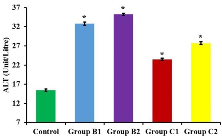
The mean value of ALT in control group (A), group B1 (25 ml Soybean oil/kg rat pellet), group B2 (35 ml Soybean oil/kg rat pellet), group C1 (25 ml Canola oil/kg rat pellet) and group C2 (35ml Canola oil/kg rat pellet) were 15.44±0.35, 32.81 ± 0.39, 35.23 ±0.26 and 23.47 ± 0.28, 27.71 ±0.38 unit/litre, respectively (Figure 1).
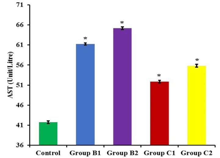
The mean value of AST in control group (A), group B1 (25ml Soybean oil/kg rat pellet), group B2 (35ml Soybean oil/kg rat pellet), group C1 (25ml Canola oil/kg rat pellet) and group C2 (35ml Canola oil/kg rat pellet) were 41.77±0.32, 61.25 ± 0.2, 65.20 ±0.33 and 51.87 ± 0.33, 55.80 ±0.36 unit/litre, respectively (Figure 2).
The ALT and AST values were increased significantly in a dose-depended aspect in both the Soybean (B1, B2) and Canola (C1, C2) oil supplemented groups comparison to the control group (A).
Gross changes in liver
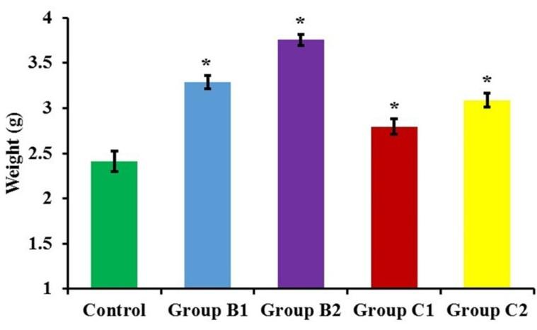
The liver of the mice of all groups was reddish in color. The mean weight of liver of groups A, B1, B2, C1 and C2 was 2.41 ± 0.11, 3.29±0.07, 3.75±0.05, 2.79±0.08 and 3.08±0.07 g, respectively (Figure 3). The mean weight of the liver was increased significantly in a dose-depended manner in both the Soybean and Canola oil supplemented groups compared to the control group.
Histopathological changes in liver
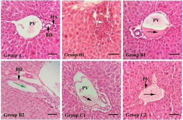
In the present study, the liver was found with normal histological architecture in the control group of mice, (Figure 4 A). Congestion was found in the portal vein of the liver of B1, C1 and C2 groups of mice (Figure 4 B1, C1 and C2). Marked lymphocytic infiltration was also found in the liver of the B1 group of mice (Figure 4 B1). The dilated bile duct was found in the liver of B2 grouped mice (Figure 4 B2).
DISCUSSION
In our present study, Swiss Albino mice were used to observe the biochemical and morphological alteration of the liver after dietary administration of Soybean and Canola oil. Serum enzymes such as ALT and AST are important liver function test to diagnose liver damage. ALT and AST enzymes are found in the heart, liver, kidney, pancreas, red blood cells and biliary ducts of the liver. AST and ALT level in the serum are used to identify body tissues particularly heart and liver is properly functioned or not. During damaging of body tissues, accessory AST and ALT are discharged into the bloodstream and increase the level of serum enzyme. Respectively, the extent of tissue damage is precisely associated with the AST and ALT level in the blood. Higher amount of AST and ALT ratio (>1.5) in acute viral hepatitis may indicate the serious condition [10]. In the biochemical study, we observed that ALT and AST values were increased significantly (p<0.05) in a dose-depended aspect in both the Soybean (B1, B2) and Canola (C1, C2) oil supplemented groups compared to control group (A). The mean weight of the liver was increased significantly in a dose-depended manner in both the Soybean and Canola oil supplemented groups compared to the control group. The present research finding is partially consistent with [1] reported that significantly higher liver weight was acquired in soybean oil supplemented groups than that of the control group.
Concerning the histopathological changes, the result of the present study revealed that there was various distortion observed in the liver of Soybean and Canola oil supplemented groups of mice. Marked lymphocytic infiltration with congestion in the portal vein was found in the liver of the B1 group of mice. The dilated bile duct was found in the liver of B2 grouped mice. The present result in soybean oil supplementation groups is partially consistent with [5] reported that distinct enlargement of central veins and portal veins with congestion and mononuclear cell infiltration was observed in the liver of soybean oil supplementation grouped rat. The present research finding in Soybean oil treated groups is not appropriate with [2] stated that the section of the liver of soybean oil supplemented group rat displayed normal histology of liver without exhibiting any observable lesions. Slight congestion of the portal vein was found in the liver of Canola oil supplemented group (C1) of mice. There was also severe congestion of the portal vein was found in the liver of Canola oil supplemented group (C2) of mice. The present research finding in Canola oil treated group (C1 and C2) is partially appropriate with [8] reported that dilated and congested hepato-portal blood vessel was found in the liver of canola oil supplemented group rat. Lipid peroxidation is a physiological mechanism that occurs in all aerobic cells. Cell membranes structural part are unsaturated fatty acids and this unsaturated fatty acids are directly associated with the lipid peroxidation by a non-enzymatic and free-radical mediated reaction chain [11]. Soybean and Canola oil are rich in unsaturated fatty acids. So more unsaturated fatty acids occur more lipid peroxidation. The toxicity of lipid peroxidation products in mammals generally involves hepatotoxicity [12]. So lipid peroxidation may be an important factor to produce histopathological lesions in Soybean and Canola oil supplemented groups of mice.
CONCLUSIONS
In the present research work, we found that ALT and AST values were increased in both the Soybean and Canola oil supplemented groups of mice comparison to the control group. Increased the level of ALT and AST values are the biomarker of liver injury and its reflection we found in the histopathological study. The different harmful histopathological lesion was found in both the Soybean and Canola oil supplemented groups of mice. The present study suggests that we should have conscious about the consumption of Soybean and Canola oil. Though oils and fats are detrimental to health but to evaluate the toxicity of Soybean and Canola oil, further studies with more laboratory animals, more duration and some other organ histopathology may be conducted.
ACKNOWLEDGEMENT
The authors extend their appreciation to the Ministry of Science and Technology, Bangladesh (MoST; Project no. BS 41/52/2018-19) for funding the research works.
AUTHOR CONTRIBUTIONS
MRI designed the experiment. MAS performed the experiments; MAS analyzed the data and wrote the draft, MRI and ZH critically revised the manuscript.
CONFLICTS OF INTEREST
The author declares that no conflict of interest exists.
References
- [1]Ahmad N, Majumder S, Miah MA, Uddin MJ. Effects of edible fats and oils on the body weight gain and on weights of some selected organs in rats removing the impact of unequal feed intake. Bangl. J. Vet. Med. 2007; 5 (1 & 2): 107–110.
- [2]Hoque M, Kabir M, Hasan M, Rahman M, Rashid M, Ruba T. Biochemical and pathological effects of palm, mustard and soybean oils in rats. Bangl. J. Vet. Med. 2018; 16 (1): 107–114.
- [3]Bhattacharya S. Seeds as Herbal Drugs. In: Preedy VR, Watson RR, Patel VB (ed). Nuts & Seeds in Health and Disease Prevention. Elsevier Inc: USA, 2011, pp 15-24.
- [4]Abdel-Misih SR, Bloomston M. Liver Anatomy. Surg Clin North Am. 2010; 90(4):643–653.
- [5]Kosif R, Yilmaz F, Evrendilek GA, Diramali M. Histopathological effects of Aloe barbadensis and soybean oil on rat liver. Int. J. Morphol. 2010; 28 (4):1101-1106.
- [6]Deol P, Evans JR, Dhahbi J, Chellappa K, Han DS, Spindler S. Soybean oil is more obesogenic and diabetogenic than coconut oil and fructose in mouse: potential role for the liver. PloS one. 2015; 10 (7): e0132672.
- [7]Henkel J, Alfine E, Saín J, Jöhrens K, Weber D, Castro J. Soybean oil-derived poly-unsaturated fatty acids enhance liver damage in NAFLD induced by dietary cholesterol. Nutrients. 2018; 10 (9): 1326.
- [8]El-Reffaei WHM, El-Sebeay AS, Ragheb EM, El-Ghandour HMA, Badr SEA. Effect of deep-fat frying on canola oil, palmolein and sunflower oil blends: b) biologicaland nutritional studies. J. Food and Dairy Sci., Mansoura Univ. 2016; 7(2): 81 -96.
- [9]Jannat N, Sultana N, Jahan MR, Islam MR. Long term administration of gentamicin affects hemato-biochemical parameters and liver archiytecture of Swiss Albino Mice. J Adv Biotechnol Exp Ther. 2018; 1 (2): 29-35.
- [10]Hasan KMM, Tamanna N, Haque MA. Biochemical and histopathological profiling of Wistar rat treated with Brassica napus as a supplementary feed. Food Sci Hum Wellness. 2018; 7(1): 77–82.
- [11]Marisa R, Boveris A, Semprine J. Lipid Peroxidation: Chemical Mechanism, Biological Implications and Analytical Determination. In: Catala A (ed). Lipid Peroxidation. In tech: Rijeka, Croatia, 2012, 03-30.
- [12]Boveris A, Repetto MG, Bustamante J, Boveris AD, Valdez LB, The concept of oxidative stress in pathology. In: Álvarez S and Evelson P (ed). Free Radical Pathophysiology. Transworld Research Network: Kerala, India, 2008, 1-17.