Epidemiological and hematological aspects of idiopathic diarrhea in dairy cattle at Sirajganj district of Bangladesh
Abstract
Cattle diarrhea poses a significant risk to the global livestock sector, leading to considerable economic setbacks and impacting the well-being of animals. This research aimed to assess the clinical and hematological changes associated with idiopathic diarrhea in cattle in the Baghabari area of the Sirajganj District of Bangladesh. The study involved surveying dairy cattle to record their case history, followed by the collection and measurement of hematological indices in those animals suffering from idiopathic diarrhea. The survey identified 39 diarrheic crossbred cattle (2 males and 37 females), ranging in age from 1 month to 11 years. The findings revealed a higher prevalence of diarrheal cases in cows (94.87%) compared to oxen (5.13%), particularly among pregnant cows (64.86%). The most common historical observations included repeat breeding (81.08%), abortion (78.38%), decreased milk production (43.24%), fever (15.64%), and congenital defects (17.95%) in diarrheic cattle. Hematological analyses indicated a significant increase in lymphocytes and immunoglobulin levels, while eosinophil, basophil, and red blood cell distribution width were notably lower in diarrheic cattle compared to healthy ones. However, other hematological parameters remained unaffected. In summary, the study suggests that bovine diarrhea may influence hematological profiles and body physiology, with the extent depending on the disease’s etiology and complexity.
INTRODUCTION
Bangladesh boasts one of the highest cattle densities, with 145 large ruminants per square kilometer (Sq. Km.), surpassing densities in India (90/Sq. Km.), Ethiopia (30/Sq. Km.), and Brazil (20/Sq. Km) [1]. The livestock sector contributes approximately 3% to the agricultural gross domestic product, supporting around 15% of the nation's workforce [2]. Given Bangladesh's agricultural focus and the dynamic potential of this emerging agricultural sub-sector, there is a need for adequate policy attention to enhance animal health and production. A significant challenge in cattle farming in this region is bovine diarrhea, considered the primary cause of growth retardation and neonatal mortality [3]. Bovine diarrhea poses a substantial threat to the global livestock industry, causing significant economic losses and impacting animal welfare [4].
The multifactorial nature of this ailment, characterized by a myriad of infectious agents, environmental factors, and host-related variables, presents a formidable challenge for effective prevention and control [5,6]. As the prevalence of bovine diarrhea continues to escalate, understanding the intricate interplay between various contributing factors becomes paramount for devising targeted interventions. Cattle diarrhea is attributed to both infectious and non-infectious factors. Various infectious enteric pathogens such as bacteria, viruses, and protozoa play leading roles in causing gastroenteritis in calves, resulting in significant morbidity and mortality [7,8]. The prevalence of each infectious agent and diarrheal case can vary depending on several factors, notably the geographical location of the farms, management practices on the farm, size of the herd, etc. [4].
Hemato-biochemical indices serve as critical indicators for diagnosing diarrheal diseases [9,10]. However, in Bangladesh, a significant number of diarrheal cases are often managed with antibiotics and other medications without a targeted identification of the underlying cause or the implementation of hematological tests. The fundamental aspect of the laboratory diagnosis of bovine diarrhea involves measuring hematological parameters such as levels of red blood cells (RBC), white blood cells (WBC), platelet (PLT), neutrophil (NEUT), lymphocyte (LYM), eosinophil (EO), basophil (BASO), monocyte (MONO), packed cell volume (PCV), immunoglobulin (IG), etc. [11,12]. Key hematological attributes associated with cattle diarrhea include higher PCV, neutrophilia, leukopenia, lymphopenia, etc. [13]. Despite this, there is a scarcity of comprehensive clinical and hematological information on idiopathic diarrhea in cattle in Bangladesh. Addressing this gap in scientific knowledge, this report aims to conduct a robust analysis of hematological indices in cattle afflicted with idiopathic diarrhea in the Baghabari area within the Sirajganj District of Bangladesh.
MATERIALS AND METHODS
Ethical approval
Ethical clearance for the handling of animals and experimental procedures was obtained from the institutional ethical committee of Bangladesh Agricultural University, Mymensingh 2202, Bangladesh (AWEEC/2023(64)).
Study location
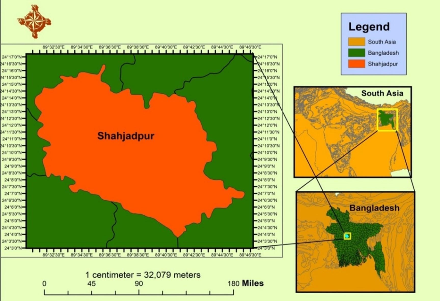
A study was conducted in the Baghabari area of Sirajganj District, Bangladesh, to examine the blood profiles of a high-yielding breed of cattle affected by idiopathic diarrhea (Figure 1).
Our investigation identified 39 affected cattle, ranging in age from 01 month to 11 years. These animals exhibited diarrhea as a prevalent symptom, along with additional clinical observations, including fever, reduced milk production, abortion, repeat breeding, and congenital defects.
Sample collection
With the owner's prior consent, 2 ml of blood sample was collected from each cattle via jugular venipuncture using sterile techniques. The collected blood samples were immediately transferred to vacutainer tubes with anticoagulant (EDTA). Blood samples were also collected from five healthy cattle to compare dynamic changes in blood profiles between healthy and diarrheic individuals.
Hematological examinations
The collected blood samples were transported to the lab in an icebox and analyzed using an auto hemo-analyzer (Zybio Hematology Analyzer Z51 VET, China). Hematological parameters including total erythrocyte count (TEC), hemoglobin (Hb), total leukocyte count (TLC), PLT, NEUT, LYM, MONO, EO, BASO, PCV, mean corpuscular volume (MCV), mean corpuscular hemoglobin (MCH), mean corpuscular hemoglobin concentration (MCHC), red blood cell distribution width (RDW-CV and RDW-SD), platelet distribution width (PDW), mean platelet volume (MPV), platelet-large cell ratio (P-LCR), procalcitonin test (PCT), and IG were analyzed using Zybio Hematology Analyzer Z51 VET, China, following the manufacturer's instructions.
Statistical analysis
All the obtained data were analyzed using IBM SPSS (version 22). Levene’s Test assessed the homogeneity of the dataset. Comparisons of blood profiles between healthy and diarrheic cattle were conducted using a paired sample t-test. The data are presented as mean ± standard error of the mean (SEM).
RESULTS
Clinical findings in diarrheic cattle
All 39 cattle with diarrhea underwent artificial insemination (AI) (Table 1). The findings indicate a higher prevalence of non-specific diarrhea in cows (94.87%) compared to oxen (5.13%). Among the 37 high-yielding dairy cows, 24 (64.86%) were at various stages of pregnancy (Table 1). Regarding additional clinical symptoms, the diarrheic cattle exhibited fever, reduced milk production, repeat breeding syndrome, a history of abortion, and congenital defects in 25.64%, 43.24%, 81.08%, 78.38%, and 17.95%, respectively.
Table 1. Clinical findings in diarrheic cattle.
Effect of idiopathic diarrhea on red blood cells and platelet counts
The analysis of RBC counts showed that TEC, PCV, MCV, MCH, MCHC, and RDW-CV were marginally increased in diarrheic cattle compared to healthy animals, but the differences were not statistically significant (P > 0.05) (Table 2 and Figure 2). However, the RDW-SD value was significantly decreased (P < 0.05) in diarrheic cattle compared to healthy animals (Table 2). Additionally, the PLT count tended to decrease (P > 0.05) in diarrheic cattle compared to healthy ones (Figure 2). Interestingly, the PDW, MPV, and P-LCR values were non-significantly increased in diarrheic cattle compared to healthy animals (Table 2).
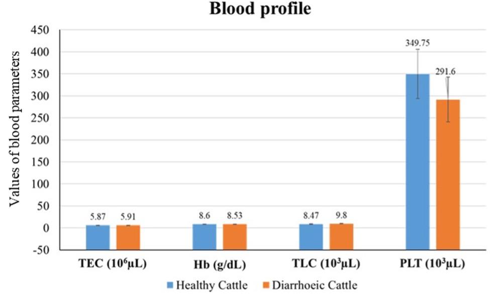
Table 2. Dynamics of hematological parameters in healthy and diarrheic cattle.
Effect of idiopathic diarrhea on white blood cells and differential leucocyte counts
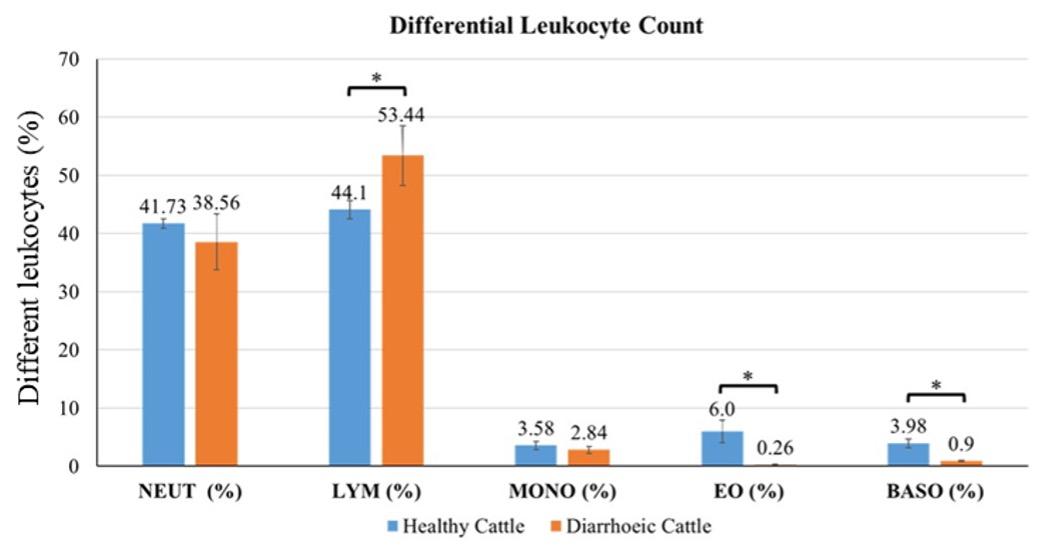
The TLC was slightly increased in diarrheic cattle compared to healthy individuals (Figure 2). The findings of the differential leukocyte count are shown in Figure 3. There was a non-significant reduction (P > 0.05) in blood NEUT and MONO levels, while EO and BASO levels decreased significantly (P < 0.05) in diarrheic cattle. On the contrary, LYM levels substantially increased (P < 0.05) in diarrheic cattle (Figure 3).
Effect of idiopathic diarrhea on inflammatory biomarkers
The PCT value increased non-significantly (P > 0.05) in diarrheic cattle compared to healthy cattle. Additionally, the IG level was significantly higher (P < 0.05) in diarrheic cattle compared to healthy individuals (Table 3).
Table 3. Dynamics of inflammatory biomarkers in healthy and diarrheic cattle.
DISCUSSION
Cattle diarrhea can result from various factors, including management practices, balantidiasis infection, dietary-related diarrhea infection, chronic salmonellosis infection, copper deficiency, Johne’s disease, chronic peritonitis, and infections with amphistomiasis or of idiopathic origin [4,5,6]. It was observed that a majority of cattle experiencing diarrhea were female, particularly pregnant cows. These animals exhibited additional clinical symptoms such as fever, reduced milk production, abortion, and repeat breeding. Pregnant and lactating cows are generally more vulnerable to various infections or management-related conditions, making them more susceptible to the development of [14]. Semisolid to watery feces with increased body temperature are common clinical findings in diarrheic cattle [9,10]. Other clinical observations in diarrheic cattle include anorexia, staggering movement, sunken eyes, mild to moderate dehydration, dullness, pale mucous membranes, and decreased milk production in lactating cows [4,10]. Body temperature could be influenced by the duration and severity of diarrhea, with an increase in temperature in the early stage and a decrease in severe cases of diarrhea [15]. Various causal agents of diarrhea in cattle, such as the bovine viral diarrhea virus, are also linked to diverse reproductive disorders like repeat breeding and abortion, justifying our current findings [16,17]. Diarrhea stands as a significant health concern in cattle, and recent research indicates a connection between hematological indices and the origins of diarrhea in these animals [6]. Consequently, hematological indices can offer valuable insights and serve as a crucial tool for the diagnosis and prognosis of diarrheal diseases in cattle [18]. TEC and Hb are important indicators for diagnosing anemic conditions. In the present study, no significant difference in TEC was found, which is in line with the previous study [19]. Nevertheless, our results are in contrast to certain other studies that reported reduced levels of TEC and Hb in cattle with diarrhea [7,10]. The levels of TEC and Hb might be linked to the etiological cause of diarrhea, stage, and chronicity of the disease [18,20]. The mean values of TLC and PLT did not exhibit a significant difference between healthy and diarrheic cattle, although the PLT level was observed to be lower in the diarrheic group. A decrease in platelet count commonly occurs in instances of severe acute viral illnesses, although this reduction in platelets doesn't consistently lead to noticeable bleeding, as confirmed by our results [21,22]. The etiology of thrombocytopenia remains unclear, but potential factors include megakaryocyte necrosis, diminished thrombocyte production by megakaryocytes, heightened peripheral thrombocyte consumption, and functional abnormalities in thrombocytes [23]. Additionally, another study investigated the direct correlation between the onset of thrombocytopenia and bone marrow infection with bovine viral diarrhea virus (BVDV) [24]. Their study revealed the presence of BVDV in all cellular components of the bone marrow, including megakaryocytes. The TLC level, serving as an indicator of septicemia, suggests a potential association between diarrhea and management practices or environmental factors in our study. Previous research has indicated a decline in TLC levels in cattle infected with bovine viral diarrhea while it increases in bacterial infections like E. coli [9, 25]. The present investigation indicated that the blood coagulation index (PLT volume) dropped non-significantly while the PDW, MPV, and P-LCR values increased in diarrheic cattle. Similar findings were reported in cattle infected with bovine viral diarrhea [25, 26]. These alterations in the haemogram suggest that diarrhea may shorten the life span of PLT.
The differential leukocyte count showed increased LYM levels and decreased EO and BASO levels, while the NEUT and MONO levels remained unaffected in the diarrheic cattle. However, all these values were within the normal range, except for EO, which was found below the normal [27]. The decreased EO level in diarrheic cattle might be associated with malnutrition or inflammatory stress [25]. Increased DLC accompanied by neutrophilia suggests diarrhea resulting from infectious agents of different origins [9,27]. An earlier study observed a noteworthy correlation between the temporal progression of innate immune responses and the viral load in the context of acute feline immunodeficiency viral infection [28]. This infection is marked by a reduction in neutrophil count and an increase in large granular lymphocyte (LGL) levels.
Our findings indicated a notable rise in LYM count and a reduction in neutrophil, eosinophil, and basophil counts in diarrheic cattle compared to their healthy counterparts. These results align with the findings reported by another study where consistently elevated LYM numbers and decreased eosinophil and basophil counts were observed in peripheral blood during bovine viral diarrheal infection [29]. Hence, the changes observed in the distribution of various white blood cell populations in our study suggest that viral infections could likely be the underlying cause of diarrhea in cattle.
The mean PCV value in diarrheic cattle is quite ambiguous. Some studies have reported decreased PCV, while many others have reported increased PCV in infectious diarrhea [7,9,19]. The elevated PCV in affected cattle might be attributed to the hemoconcentration associated with dehydration and hypovolemia resulting from excessive fluid loss [27]. However, in the current investigation, the levels of PCV, MCV, MCH, MCHC, RDW-CV, and PCT were found to be similar in healthy and diarrheic cattle. A previous study reported substantially increased MCV and MCH values in colibacillosis-infected diarrheic calves [9]. Conversely, another study reported decreased MCV, MCH, and MCHC values [7]. However, these parameter levels may be related to the underlying cause and severity of the diarrhea. The RDW-SD value was significantly lower in diarrheic cattle, indicating a very minimal difference in red blood cell size. Nevertheless, a low RDW value is an indicator of macrocytic anemia [30]. The IG level is an excellent indicator of the immune status of an individual and might be affected by multiple factors such as nutritional status, stress levels, infection, chronic health conditions, etc. [31]. In the current study, the IG level increased significantly in the diarrheic cattle, which might be due to the presence of idiopathic subclinical infection [32]. However, the actual causes of diarrhea in dairy cattle in the study area could not be determined, which is the main limitation of this study and should be investigated in the future for better and specific treatment and management.
CONCLUSION
Diarrheal infection in cattle was exhibited through different clinical findings, with significant changes in hematological parameters such as LYM, PLT, EO, and BASO in diarrheic animals compared to healthy controls. These findings will provide valuable diagnostic indices for the cattle industry as well as for future research, which may help to control diarrheal disease. However, this is the first report so far, on the hematological profile analysis in the high-yielding cattle in Bangladesh and further study is needed to investigate the disease in detail.
ACKNOWLEDGMENT
We would like to thank Bangladesh Agricultural University, Mymensingh 2202, Bangladesh, for providing the research grants (Project No. 2021/1380/BAU) to conduct this investigation.
AUTHORS CONTRIBUTION
MGH conceptualized and supervised the experiment. SMNH and MAU collected the case history and blood samples. MGH, SMNH, AM and MAU conducted the experiments. MRI analyzed the data, interpreted the results, and drafted the manuscript. MJM, MA, NJ, MEJB, MGH and SA critically revised and edited the manuscript. All authors gave final approval and agreed to be accountable for all aspects of work in ensuring that questions relating to the accuracy or integrity of any part of the work.
CONFLICTS OF INTEREST
There is no conflict of interest among the authors.
References
- [1]Pallab M, Ullah S, et al. A cross sectional study of several diseases in cattle at chandanaish upazilla of chittagong district, bangladesh. Sci J Vet Adv. 2012;1:28-32.
- [2]Haider N, Rahman M, et al. Identification and epidemiology of a rare hobi‐like pestivirus strain in b angladesh. Transbound Emerg Dis. 2014;61:193-8.
- [3]Rahman MM, Kabir A, et al. The dynamics of bovine viral diarrhea virus (bvdv) infection and possible impacts on cattle reproduction. Bangladesh Livest J. 2017;3:1-6.
- [4]Cho Y-i, Yoon K-J. An overview of calf diarrhea-infectious etiology, diagnosis, and intervention. J Vet Sci. 2014;15:1-17.
- [5]Blanchard PC. Diagnostics of dairy and beef cattle diarrhea. Vet Clin North Am Food Anim. 2012;28:443-64.
- [6]Hassan N, Randhawa CS, et al. Evaluating the hemato-biochemical indices in relation to the different etiologies of chronic diarrhea in dairy cattle and buffalo. Comp Clin Path. 2022;31:585-95.
- [7]Kumar S, Jakhar K. Haematological, biochemical and oxidative stress parameter as prognostic indicators in calf diarrhoea. J Pharm Innov. 2020;9:10-3.
- [8]Singh M, Gupta V, et al. A study on alteration in haemato-biochemical parameters in colibacillosis affected calves. Int j adv res. 2014;2:746-50.
- [9]Nayak T, Singh A, et al. A study on alteration in clinico-haematological parameters in colibacillosis affected diarrhoeic cattle calves. Int J Chem Stud. 2019;7:1559-62.
- [10]Shehta A, El-Zahar H, et al. Clinical, hematological and some biochemical alterations during diarrhea in friesian calves naturally infected with e. Coli and salmonella. Beni-Suef univ j basic appl sci. 2022;11:1-8.
- [11]Mohan G, Kumar A, et al. Effect of rumen fermentative disorders on physiological parameters in buffaloes. Int J Vet Sci. 2015;4:10-4.
- [12]Youssef MA, El-Ashker MR, et al. Hematological and serum biochemical alterations in buffalo with some digestive disorders. Comp Clin Path. 2017;26:1033-9.
- [13]Alsaad K, Al-Obaidi Q, et al. Clinical, haematological and coagulation studies of bovine viral diarrhoea in local iraqi calves. Bulg J Vet Med. 2012;15.
- [14]Kelling CL, Topliff CL. Bovine maternal, fetal and neonatal responses to bovine viral diarrhea virus infections. Biologicals. 2013;41:20-5.
- [15]Torche S, Boussena S, et al. Physiopathology of diarrhea in young calves: Clinical signs and metabolic disturbances. J New Sci. 2020;76.
- [16]Walz PH, Givens MD, et al. Evaluation of reproductive protection against bovine viral diarrhea virus and bovine herpesvirus-1 afforded by annual revaccination with modified-live viral or combination modified-live/killed viral vaccines after primary vaccination with modified-live viral vaccine. Vaccine. 2017;35:1046-54.
- [17]Asmare K, Sibhat B, et al. Serological evidence of bovine herpesvirus-1, bovine viral diarrhea virus and schmallenberg virus infections in relation to reproductive disorders in dairy cattle in ethiopia. Acta Trop. 2018;178:236-41.
- [18]Chae J-B, Ku J-Y, et al. Hematology, serum biochemistry, and acute phase proteins in hanwoo (bos taurus coreanae) calves with diarrhea. J Vet Clin. 2022;39:342-52.
- [19]Jaiswal M, Shukla P, et al. Clinical score and haemato-biochemical alterations in acute diarrhoea in calves. J Pharm Innov. 2019;8:529-32.
- [20]Villa L, Gazzonis AL, et al. Exploring alterations in hematological and biochemical parameters, enzyme activities and serum cortisol in besnoitia besnoiti naturally infected dairy cattle. Parasites & Vectors. 2021;14:1-13.
- [21]Rebhun WC, French TW, et al. Thrombocytopenia associated with acute bovine virus diarrhea infection in cattle J Vet Intern Med. 1989;3:42-6.
- [22]Corapi WV, Elliott RD, et al. Thrombocytopenia and hemorrhages in veal calves infected with bovine viral diarrhea virus. J Am Vet Med Assoc. 1990;196:590-6.
- [23]Walz PH, Steficek BA, et al. Effect of experimentally induced type ii bovine viral diarrhea virus infection on platelet function in calves. Am J Vet Res.1999;60:1396-5.
- [24]Spagnuolo M, Kennedy S, et al. Bovine viral diarrhoea virus infection in bone marrow of experimentally infected calves. J Comp Pathol. 1997;116:97-100.
- [25]Hasan S, Alsaad KM. Evaluation of clinical, hematological, blood coagulation and some biochemical parameter changes in clinically infected cattle with bovine viral diarrhea. J Agric Vet Sci. 2018;11:64-70.
- [26]Radwińska J. Effect of the bvd-md virus on coagulation and fibrinolytic systems in dairy cows. Bull Vet Inst Pulawy. 2010;54:293-8.
- [27]Barua SR, Md Rakib T, et al. Hematological and serological changes in neonatal diarrheic calves infected with bovine rotavirus. Multidisciplinary Advances in Veterinary Science. 2018;2:356-66.
- [28]Sprague W, TerWee J, et al. Temporal association of large granular lymphocytosis, neutropenia, proviral load, and fasl mrna in cats with acute feline immunodeficiency virus infection. Vet Immunol Immunopathol. 2010;134:115-21.
- [29]Chowdhury M, Afrin F, et al. Prevalence and haematological parameters for bovine viral diarrhoea (bvd) in south bengal areas in bangladesh. Bangladesh Veterinarian. 2015;32:48-54.
- [30]Herman N, Trumel C, et al. Hematology reference intervals for adult cows in france using the sysmex xt-2000iv analyzer. J Vet Diagn Invest. 2018;30:678-87.
- [31]Chung JJ, Rayburn MC, et al. Randomized controlled clinical trial on the effect of oral immunoglobulin supplementation on neonatal dairy calves with diarrhea. J Vet Intern Med. 2019;33:1807-13.
- [32]Rocha T, Silva F, et al. Serum concentrations of acute phase proteins and immunoglobulins of calves with rotavirus diarrhea. Arq Bras Med Vet Zootec. 2016;68:865-72.