Prophylactic effects of vitamin E and coriander (Coriandrum sativum) extract on lead-induced testicular damage in Swiss albino mice
Abstract
Lead toxicity is the vital issue in the developing countries that causes serious health hazards in animals and humans. Prophylactic effects of vitamin E and coriander (Coriandrum sativum) extract on lead-induced testicular damage in Swiss albino mice were investigated in the present study by gross and histological studies in five groups (control, lead intoxicated, vitamin E-treated, coriander-treated, vitamin E and coriander-treated). Treatment was done for 42 days. In the present study, weight and length of left and right testes were significantly (p<0.05) reduced in lead-intoxicated mice in comparison to the control group. Lead acetate was found to cause separation of primary spermatocyte from spermatogonia, morphological changes of seminiferous tubule and irregular arrangement of spermatogenic cells in the seminiferous tubules of testis. Vitamin E and coriander extract were found effective in the treatment of lead intoxicated mice and testes histology were found normal. Combined antioxidative action of vitamin E and corriander extract was also found effective in the treatment of lead intoxicated mice. The present investigation may serve as baseline data about adverse effects of lead toxicity and efficacy of vitamin E and coriander extract against lead toxicity. Further research need to be done to isolate and purify the active principle involved in the antioxidant activity of this plant.
INTRODUCTION
The humans and animals are exposed to various types of environmental contaminants at various stages of their life, majority of them are hazardous to health. Lead is one of them. Lead is considered the most common occupational and environmental toxicants and it has stern potential health hazards to humans (especially children) and animals. In Bangladesh, livestock are affected by lead toxicity. Mainly soft tissues like liver, kidney and testis are affected seriously. Most lead-associated renal effects or toxicity are a result of the ongoing chronic or current high acute exposure. Lead exposure causes progressive vascular, tubular and interstitial testicular damage [1].
Many researchers have used a wide variety of antioxidants including vitamin C [2] and vitamin E [3-4] to prevent the incidence and to lessen oxidative stress in tissues. Vitamin E minimizes the impact of lead induced reduction in sperm count in Swiss mice. Similarly, Mishra and Acharya [5] also observed that vitamin E reduced the impact of lead induced reduction in sperm count in Swiss mice. Vitamin E was found to exhibit a protective effect on the testis of rats [6]. They also stated that vitamin E functions as a free radical scavenger, scavenging superoxide, hydrogen peroxide and hydroxyl radicals. Similarly, other group also reported that Vitamin E has beneficial effects to protect sperm DNA from oxidative stress of free radicals and to improve fertility [7]. It is a chief chain-breaking antioxidant in the sperm membranes. Adding vitamin E in diet enhanced the dynamism of some antioxidant enzymes, decreased nitric oxide content and lipid peroxidation products in the testis of Boer goat [8].
The seed of coriander is one of the most valuable spices in the world. It is regularly used by South Asian kitchen. In addition to its cooking value, coriander is popular for its wide range of healing properties. It assists to remove toxic mineral residue such as mercury and lead from the body through the urine or faces. Coriander (Coriandrum sativum) promotes SOD, CAT functions and GSH content and decreases LPO level in lead induced mice tissues. The antioxidant property of coriander extracts could be directly related to both the scavenging function against ROS and elevation of antioxidant make up. A study shown that the formation of lipid peroxides declined and the activities of antioxidant enzymes (catalase, glutathione peroxidase) increased in rats treated by coriander extracts [9].
This study was designed to investigate the effects of lead toxicity on testis of mice and possible curable effects produced by vitamin E and extract of coriander seeds supplementation.
MATERIALS AND METHODS
Chemicals
The study was conducted in the Department of Anatomy and Histology, Faculty of Veterinary Science, Bangladesh Agricultural University, Mymensing-2202. Vitamin E was purchased from commercial source. Coriander extract was prepared in the Department of Pharmacology, Faculty of Veterinary Science, Bangladesh Agricultural University, Mymensingh.
Animals and Treatments
The experimental Swiss albino mice (male) were collected from Department of Pharmacy, Jahangirnagar University, Dhaka. Collected mice were 6 weeks of age and about 26-28 grams at the time of collection. All mice were managed and raised under confinement as an intensive system. Mice were kept cages at room temperature. The mice were fed with feed and water ad libitum. All experimental protocols were approved by the Animal Welfare and Ethical Committee (order no. sha 1/444/edu) Faculty of Veterinary Science, Bangladesh Agricultural University.
After 7 days, mice were divided into different groups according to the experimental design. At first there was two groups- Group A: Control group (10 mice) and Group B: Lead intoxicated group (25 mice). Control group was received only normal water and feed. Lead intoxicated group was treated with 60 mg lead acetate per kg body weight in every day orally for 6 weeks. After six weeks samples were collected from 5 mice of control group and 5 mice of intoxicated group. Remaining 5 mice of control group were kept as control for next 42 days. Five mice of intoxicated group were further intoxicated for next 42 days. Other 15 mice of intoxicated group were kept for treatment purpose. These mice were divided into three groups (C, D and E) each having 5 mice. Group C was treated with 150 mg vitamin E (diluted in soya oil) per kg body weight in every day orally for 6 weeks. Group D was treated with 300 mg coriander extract (diluted in distilled water) per kg body weight in every day orally for 6 weeks. Group E was treated with 150 mg vitamin E (diluted in soya oil) and 300 mg coriander extract (diluted in distilled water) per kg body weight in every day orally for 6 weeks. After completion of experiment, samples were collected from all the mice of different groups.
Gross and Histology
In gross study, parameters such as color, length and weight were taken into consideration. All kind of abnormalities were also observed. The color of testis was compared with the organs of control group by eye observation. Length of testis of different groups was measured by a graded scale. Unit of length measurement was milimeter. Weight was measured in gram by electronic balance.
After gross observation, samples were preserved in 10% formalin and Bouin’s fluid. After proper fixation, samples were processed for histological study. H & E staining protocol was applied. The details histological study was done using a light microscope.
Photomicrographs
Photographs for the present study were taken according to our previous study [10]. Necessary photomicrographs were taken with Olympus BX 51 photographic light microscope and placed for better illustration of the result.
Data Analysis
All the collected data were then analyzed using Statistical Package for the Social Sciences (SPSS) software and disrobe the results in tabular form. The chi- squared test was used for the analytical assessment. The differences were considered statistically significant when the p values were less than 0.05.
RESULTS
Gross architectural changes
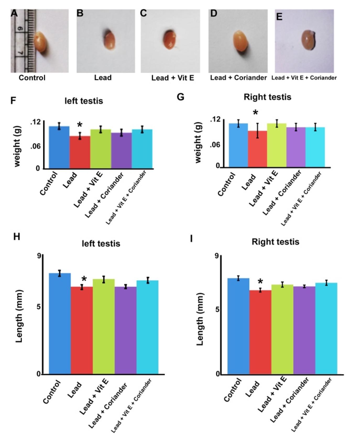
Appearance of testis was found normal in control group, vitamin E treated group, coriander extract treated group and vitamin E and coriander extract (combined) treated group. In lead intoxicated group, testis was found in reduced size (Fig. 1A-E). The mean weight of left testis in control group, intoxicated group, vitamin E treated group, coriander extract treated group and vitamin E and coriander extract (combined) treated group was 0.13 ± 0.01, 0.10 ± 0.01, 0.12 ± 0.01, 0.11 ± 0.01 and 0.12 ± 0.01 g, respectively (Fig. 1F). Weight of left testis in lead intoxicated group was significantly (p<0.05) reduced in comparison to the control group. The mean weight of right testis in control group, intoxicated group, vitamin E treated group, coriander extract treated group and vitamin E and coriander extract (combined) treated group was 0.12 ± 0.01, 0.10 ± 0.02, 0.12 ± 0.01, 0.11 ± 0.01 and 0.11 ± 0.01 g, respectively (Fig.1G). Weight of right testis in lead intoxicated group was significantly (p<0.05) reduced in comparison to the control group.
The mean length of left testis in control group, intoxicated group, vitamin E treated group, coriander extract treated group and vitamin E and coriander extract (combined) treated group was 7.59 ± 0.23, 6.57 ± 0.17, 7.14 ± 0.24, 6.58 ± 0.14 and 7.07 ± 0.20 mm, respectively (Fig. 1H). Length of left testis in lead intoxicated group was significantly (p<0.05) reduced in comparison to the control group. The mean length of right testis in control group, intoxicated group, vitamin E treated group, coriander extract treated group and vitamin E and coriander extract (combined) treated group was 7.43 ± 0.17, 6.50 ± 0.15, 6.93 ± 0.20, 6.79 ± 0.10 and 7.07 ± 0.20 mm, respectively (Fig. 1I). Length of right testis in lead intoxicated group was significantly (p<0.05) reduced in comparison to the control group.
Microscopic architectural changes
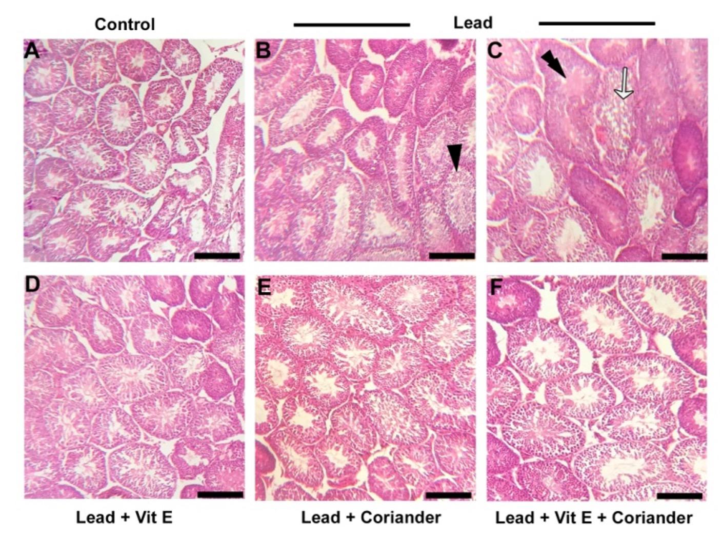
In the present study, appearance of seminiferous tubules was found normal in control group. Spermatogenic cells were present in normal pattern (Fig. 2 A). In lead intoxicated group, primary spermatocytes were found separated from spermatogonia (Fig. 2B). In addition, morphological changes of seminiferous tubules and irregular arrangement of spermatogenic cells were also observed in this group (Fig. 2 C). Seminiferous tubules were found normal in vitamin E treated group, coriander extract treated group and vitamin E and coriander extract treated group. Primary spermatocytes were not found separated from spermatogonia in these groups. Spermatogenic cells were present in normal pattern. Morphological changes were not observed in the seminiferous tubules (Fig. 2 D-F).
DISCUSSION
In the present study, the prophylactic effects of vitamin E and coriander (Coriandrum sativum) extract on lead-induced testicular damage in Swiss albino mice were investigated in detail by gross and histological studies in five groups (control, lead intoxicated, vitamin E-treated, coriander-treated, vitamin E and coriander-treated).
Testis was found in reduced size in lead intoxicated group. This is similar to the findings of other groups [11-12] who found that the weight of the testis, epididymis and accessory sex glands was significantly decreased in rats treated with lead compared to the control group. They also reported an increase in the relative weights of the testis and epididymides in rats poisoned with 8mg/Kg/day by I.P route for five weeks. The weight of the testis is largely dependent on the mass of differentiated spermatogenic cells. Hence a reduction in its weight might be due to the decreased number of germ cells and elongated spermatids [13].
In the present study, primary spermatocyte was found to be separated from spermatogonia in lead intoxicated group. Morphological change in the seminiferous tubules and irregular arrangement of spermatogenic cells were also found in this group. This result is similar or nearly similar to the findings of previous works. It has been reported that lead causes degeneration and necrosis of spermatogonia and interstitial cells and abnormal distribution of spermatozoa [14]. In addition it was showed that lead exposure caused progressive vascular, tubular and interstitial testicular damage [1]. Other group of researcher found more conspicuous degenerative changes in testicular tissues and an increase in sperm head abnormalities in mice exposed to lead acetate [15]. It has also been reported that lead is a male reproductive toxicant and lead causes morphological changes in the seminiferous tubules [16].
The seminiferous tubules were found normal in vitamin E treated group and coriander extract treated group as well as vitamin E and coriander extract (combined) treated group in the present study. This is due to antioxidative properties of vitamin E and coriander extract. However, vitamin E has many other properties, which was found to prevent the incidence and to lessen oxidative stress in tissues [3], protective effect on the testis of rats [6] primary component of the antioxidant system of the spermatozoa [17], dynamism of some antioxidant enzymes [8] and improvement of reproductive efficiency of male rats [18-19]. The coriander extract has antioxidative properties on the testis of mice exposed to lead toxicity in the present study [20]. They suggested that aqueous and ethanolic extracts of Coriandrum sativum can prevent or slow down the oxidative damage induced by lead in mice. They also reported that the coriander mediated suppression of the increased AST and ALT activities and cholesterol level suggests the possibility of the extract to give protection against hepatic, renal and testicular injury upon lead induction.
CONCLUSION
The present findings revealed that lead has detrimental effects on testis of mice. Lead was found to cause decrease weight of testis, separation of primary spermatocyte from spermatogonia, morphological changes of seminiferous tubule and irregular arrangement of spermatogenic cells in the seminiferous tubules. Treatment with vitamin E as well as coriander extract is very effective in lead intoxication. After treatment with vitamin E and coriander extract, gross and histoarchitecture of testis were found normal.
ACKNOWLEDGEMENT
The authors extend their appreciation to the Ministry of Science and Technology, Bangladesh (MoST; Project no. BS 54/58/2017-18) for funding the research works.
AUTHORS CONTRIBUTION
MRI and MAJ designed the experiment. MRI and MAJ performed the experiments; MAJ and MNI analyzed the data and wrote the draft, MRI and MZIK critically revised the manuscript.
CONFLICT OF INTEREST
The author declares that no conflict of interest exists.
References
- [1]Moniem AEA, Dkhil MA, Al-Quraishy S. Protective role of flaxseed oil against lead acetate induced oxidative stress in testes of adult rats. Afr. J. Biotechnol. 2010; 9: 7216-7223.
- [2]Hsu CP, Guo LY. Antioxidant nutrients and lead toxicity. Toxicol. 2002; 180: 33-44.
- [3]Patra RC, Rautray AK, Swarup D. Oxidative stress in lead and cadmium toxicity and its amelioration. Veterinary Medicine International p.1-9. 2011.
- [4]Alam MS, Hoque MN. Prophylactic effects of vitamin E and selenium on di (n-butyl) phthalate-induced testicular damage in prepubetral rats. J Adv Biotechnol Exp Ther. 2018; 1 (3): 65-71.
- [5]Mishra M, Acharya UR. Protective action of vitamins on the spermatogenesis in lead-treated Swiss mice. J. Trace Elements Med. Biol. 2004; 18:173-178.
- [6]Kartikeya M, Ashok A, Rakesh S. Oxidative stress & male infertility. Indian J Med Res. 2009; 129:357–367.
- [7]Tarin JJ, Brines J, Cano A. Antioxidants may protect against infertility. Hum. Reprod. 1998; 13: 1415-1416.
- [8]Hong Z, Hailing L, Hui M, Guijie Z. Effect of Vitamin E supplementation on development of reproductive organs in Boer goat. Anim. Reprod. Sci. 2009; 113: 93-101.
- [9]Chithra V, Leelamma S. Coriandrum sativum changes the levels of lipid peroxidase and activity of antioxidant enzymes in experimental animals. Ind J Biochem Biophys. 1999; 36:59-61.
- [10]Jannat N, Sultana N, Jahan MR, Islam MR. Long term administration of gentamicin affects hemato-biochemical parameters and liver archiytecture of Swiss Albino Mice. J Adv Biotechnol Exp Ther. 2018; 1 (2): 29-35.
- [11]Hamadouche NA, Sadi N, Kharoubi O, Slimani M, Aoues A.: The protective effect of vitamin E against genotoxicity of lead acetate intraperitoneal administration in male rat. Arch. Biol. Sci. Belgrade. 2013; 65:1435–1445.
- [12]Thoreux-Manlay A, Vélez de la Calle JF, Olivier MF. Impairment of testicular endocrine function after lead intoxication in the adult rat. Toxicology. 1995;100: 101-109.
- [13]Chapin RE, Harris MW, Davis BJ, Ward SM, Wilson RE, Mauney MA, Lockhart AC, Smialowicz RJ, Moser VC, Burka LT and Collins BJ. The effects of perinatal/ juvenile methoxychlor exposure on adult rat nervous, immune and reproductive system function. National Toxicology Program, NIEHS, North Carolina, USA. 1997.
- [14]Nadia AH, Sadi Nesrine, Kharoubi O, Slimani M and Aoues A. The Protective Effect of Vitamin E Against Genotoxicity of Lead Acetate Intraperitoneal Administration in Male Rat. Arch. Biol. Sci. 2913; 65(4): 1435-1445.
- [15]Gautam AK, Agarwal K, Shah BA, Kumar S, Saiyed HN. Lead induced sperm toxicity in mouse and MPG treatment. J. Environ. Biol. 2001; 22: 287–291.
- [16]Winder C. Reproductive and chromosomal effect of occupational exposure to lead on the male. Reproductive Toxicology. 1989; 3(4): 221-233.
- [17]Hsu PC, MY Liu, CC Hsu, LY Chen, YL Guo. Effects of vitamin E and/or C on reactive oxygen species-related lead toxicity in the rat sperm. Toxicology. 1998; 128:169-179.
- [18]Kutlubay R, Can E, Nancy L. Vitamin E Protection from Testicular Damage caused in traperitoneal aluminium. International J. of toxicology. 2006; 26 (4) 297-306.
- [19]Jamilah M. Pumpkin Seed Oil and Vitamin E improve Reproductive Function of male as significant by Testicular Injury. World Applied Sci. J. 2013; 23(10): 1351-1359.
- [20]Kansal L, Sharma A, Lodi S. Remedial effect of Coriandrum sativum (coriander extracts on lead induced oxidative damage in soft tissues of Swiss albino mice. International Journal of Pharmacy and Pharmaceutical Sciences. 2012; 4:729-736.